Anti-PINK1 antibody
-
概述
- 产品描述A member of the serine / threonine protein kinase family, PTEN induced putative kinase 1 (PINK1) is a tumor suppressor. PINK1 is primarily located in mitochondria, and is ubiquitously expressed in testis, skeletal muscle, and heart tissue. at lower levels in pancreas, ovary, brain, placenta, kidney, liver, prostate and small intestine. During cellular stress PINK1 protects against mitochondrial dysfunction by inducing phosphorylation mitochondrial proteins. PINK1 mutations may give rise to different autophosphorylation activity. Mutations in the PINK1 gene (PARK6) are associated with early onset Parkinson's disease, a recessive neurodegenerative disorder characterized by resting tremor, muscular rigidity, bradykinesia and postural instability. Parkinson's disease generally involves the presence of intraneuronal accumulations of aggregated proteins (Lewy bodies) in brain neurons.
- 产品名称Anti-PINK1 antibody
- 分子量63 kDa
- 种属反应性Human,Mouse
- 验证应用WB,ICC,IHC-P
- 抗体类型兔多抗
- 免疫原Recombinant protein
- 偶联Non-conjugated
-
性能
- 形态Liquid
- 浓度1 mg/mL.
- 存放说明Store at +4℃ after thawing. Aliquot store at -20℃ or -80℃. Avoid repeated freeze / thaw cycles.
- 存储缓冲液1*PBS (pH7.4), 0.2% BSA, 50% Glycerol. Preservative: 0.05% Sodium Azide.
- 亚型IgG
- 纯化方式Protein affinity purified
- 亚细胞定位Mitochondrion outer membrane. Cytoplasm.
- 其它名称BRPK antibody
FLJ27236 antibody
mitochondrial antibody
PARK 6 antibody
PARK6 antibody
Phosphatase and Tensin Homolog antibody
PINK 1 antibody
PINK1 antibody
PINK1_HUMAN antibody
Protein kinase BRPK antibody
PTEN induced putative kinase 1 antibody
PTEN induced putative kinase protein 1 antibody
PTEN-induced putative kinase protein 1 antibody
Serine/threonine kinase PINK1 mitochondrial antibody
Serine/threonine protein kinase PINK1 mitochondrial antibody
Serine/threonine-protein kinase PINK1 antibody
more
-
应用
WB: 1:500-1:1,000
ICC: 1:50-1:200
IHC-P: 1:100-1:200
-
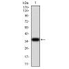
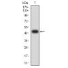
Fig1: Western blot analysis of PINK1 on different lysates using anti-PINK1 antibody at 1/200 dilution.
Positive control:
Lane 1: A431
Lane 2: Mouse testis tissue
Lane 3: Jurkat
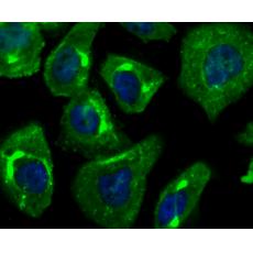
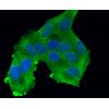
Fig2: ICC staining PINK1 in Hela cells (green). The nuclear counter stain is DAPI (blue). Cells were fixed in paraformaldehyde, permeabilised with 0.25% Triton X100/PBS.
Fig3: ICC staining PINK1 in PC-3M cells (green). The nuclear counter stain is DAPI (blue). Cells were fixed in paraformaldehyde, permeabilised with 0.25% Triton X100/PBS.
Fig4: ICC staining PINK1 in SK-Br-3 cells (green). The nuclear counter stain is DAPI (blue). Cells were fixed in paraformaldehyde, permeabilised with 0.25% Triton X100/PBS.
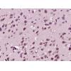
Fig5: Immunohistochemical analysis of paraffin-embedded human breast tissue using anti-PINK1 antibody. Counter stained with hematoxylin.
Fig6: Immunohistochemical analysis of paraffin-embedded human placenta tissue using anti-PINK1 antibody. Counter stained with hematoxylin.
Fig7: Immunohistochemical analysis of paraffin-embedded human stomach cancer tissue using anti-PINK1 antibody. Counter stained with hematoxylin.
特别提示:本公司的所有产品仅可用于科研实验,严禁用于临床医疗及其他非科研用途!