Anti-FCER1A antibody [A9-F2]
-
概述
- 产品描述IgE Fc Receptor I binds to the Fc region of immunoglobulin e chain with high affinity, and is responsible for initiating the allergic response. Binding of allergen to receptor-bound IgE leads to cell activation and the release of mediators such as histamines, responsible for the manifestations of allergy. IgE Fc Receptor I also induces the secretion of important lymphokines, effectors of the hypersensitivity response. Receptor I is a tetramer of a heavily glycosylated α chain (Fc ε RIα), β chain and two disulfide linked γ chains. Fc ε RIα is exposed to the outer surface of the cell and contains the IgE binding site. Expression of IgE Fc RI mRNA appears to be highly specific and has been identified in mast cells and IL-3 dependent myeloid-monocyte precursor. Alternative splicing of the genomic transcript for the a chain has also been identified.
- 产品名称Anti-FCER1A antibody [A9-F2]
- 种属反应性Human
- 验证应用WB,FC
- 抗体类型小鼠单抗
- 免疫原Recombinant protein
- 偶联Non-conjugated
-
性能
- 形态Liquid
- 浓度2 mg/mL.
- 存放说明Store at +4℃ after thawing. Aliquot store at -20℃ or -80℃. Avoid repeated freeze / thaw cycles.
- 存储缓冲液1*TBS (pH7.4), 1%BSA, Preservative: 0.05% Sodium Azide.
- 亚型IgG1
- 纯化方式Protein A purified.
- 亚细胞定位Cell membrane.
- 其它名称
- Fc epsilon RI alpha antibody
- Fc epsilon RI alpha chain antibody
- Fc epsilon RI alpha-chain antibody
more
-
应用
WB: 1:500-1:1,000
FC: 1:100-1:200
-
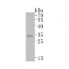
Fig1: Western blot analysis of FCER1A on human FCER1A recombinant protein using anti-FCER1A antibody at 1/1,000 dilution.
Fig2: Western blot analysis of FCER1A on different cell lysate using anti-FCER1A antibody at 1/1,000 dilution.
Positive control: Line1:SW620 Line2:A549 Line3:A431
Fig3: Flow cytometric analysis of HEK293 cells with FCER1A antibody at 1/100 dilution (green) compared with an unlabelled control (cells without incubation with primary antibody; red).
特别提示:本公司的所有产品仅可用于科研实验,严禁用于临床医疗及其他非科研用途!


![Anti-FCER1A antibody [A9-F2]](images/202012/goods_img/92767_G_1608032377590.jpg)


