Anti-EGFR antibody
-
概述
- 产品描述The EGF receptor family comprises several related receptor tyrosine kinases that are frequently overexpressed in a variety of carcinomas. Members of this receptor family include EGFR (HER1), Neu (ErbB-2, HER2), ErbB-3 (HER3) and ErbB-4 (HER4), which form either homodimers or heterodimers upon ligand binding. Exons in the EGFR gene product are frequently either deleted or duplicated to produce deletion mutants (DM) or tandem duplication mutants (TDM), respectively, which are detected at various molecular weights. EGFR binds several ligands, including epidermal growth factor (EGF), transforming growth factor α (TGFα), Amphiregulin and heparin binding-EGF (HB-EGF). Ligand binding promotes the internalization of EGFR via Clathrin-coated pits and its subsequent degradation in response to its intrinsic tyrosine kinase. EGFR is involved in organ morphogenesis and maintenance and repair of tissues, but upregulation of EGFR is associated with tumor progression. The oncogenic effects of EGFR include initiation of DNA synthesis, enhanced cell growth, invasion and metastasis. Abrogation of EGFR results in cell cycle arrest, apoptosis or dedifferentiation of cancer cells, suggesting that EGFR may be an effective therapeutic target.
- 产品名称Anti-EGFR antibody
- 分子量175 kDa
- 种属反应性Human
- 验证应用WB,ICC,IHC-P
- 抗体类型兔多抗
- 免疫原Peptide
- 偶联Non-conjugated
-
性能
- 形态Liquid
- 浓度1 mg/mL.
- 存放说明Store at +4℃ after thawing. Aliquot store at -20℃ or -80℃. Avoid repeated freeze / thaw cycles.
- 存储缓冲液1*PBS (pH7.4), 0.2% BSA, 40% Glycerol. Preservative: 0.05% Sodium Azide.
- 亚型IgG
- 纯化方式Peptide affinity purified
- 亚细胞定位Secreted and Cell membrane. Endoplasmic reticulum membrane.
- 其它名称
- Avian erythroblastic leukemia viral (v erb b) oncogene homolog antibody
- Cell growth inhibiting protein 40 antibody
- Cell proliferation inducing protein 61 antibody
more
-
应用
WB: 1:500-1:1000
ICC: 1:50-1:200
IHC-P: 1:50-1:200
-
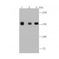
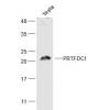
Fig1: Western blot analysis of EGFR on different cell lysate using anti-EGFR antibody at 1/1,000 dilution.
Positive control:
Lane 1: Hela
Lane 2: HUVEC
Lane 3: A431
Fig2: ICC staining EGFR in A431 cells (green). The nuclear counter stain is DAPI (blue). Cells were fixed in paraformaldehyde, permeabilised with 0.25% Triton X100/PBS.
Fig3: Immunohistochemical analysis of paraffin-embedded human tonsil tissue using anti-EGFR antibody. Counter stained with hematoxylin.
Fig4: Immunohistochemical analysis of paraffin-embedded human lung cancer tissue using anti-EGFR antibody. Counter stained with hematoxylin.
Fig5: Immunohistochemical analysis of paraffin-embedded human liver tissue using anti-EGFR antibody. Counter stained with hematoxylin.
Fig6: Immunohistochemical analysis of paraffin-embedded human kidney tissue using anti-EGFR antibody. Counter stained with hematoxylin.
Fig7: Immunohistochemical analysis of paraffin-embedded 2 kidney tissue using anti-EGFR antibody. Counter stained with hematoxylin.
特别提示:本公司的所有产品仅可用于科研实验,严禁用于临床医疗及其他非科研用途!