Anti-TrkA antibody
-
概述
- 产品描述The Trk proto-oncogene encodes a tyrosine protein kinase, Trk A, also designated Trk gp140, that serves as a receptor for certain neurotrophic factors including nerve growth factor (NGF) and neurotrophin-3 (NT-3). Trk B is a tyrosine kinase gene highly related to Trk A. Trk B expression is confined to tissues within the central and peripheral nervous systems. The brain-derived neurotrophic factor (BDNF) and NT-3, but not NGF, can induce rapid phosphorylation on Tyrosine of Trk B gp145, one of the receptors encoded by Trk B, although BDNF elicits a response at least two orders of magnitude greater than NT-3. Thus it appears that Trk B gp145 may represent a neurotrophic receptor for BDNF and NT-3. The third member of the Trk family of tyrosine kinases, Trk C, encodes a protein designated Trk C gp145 that is preferentially expressed in brain tissue, is equally related to Trk A and Trk B and is a functional receptor for NT-3.
- 产品名称Anti-TrkA antibody
- 分子量Predicted band size 87 kDa
- 种属反应性Human,Mouse,Rat
- 验证应用IHC-P,FC,WB,ICC
- 抗体类型兔多抗
- 免疫原Recombinant protein
- 偶联Non-conjugated
-
性能
- 形态Liquid
- 浓度1 mg/mL.
- 存放说明Store at +4℃ after thawing. Aliquot store at -20℃ or -80℃. Avoid repeated freeze / thaw cycles.
- 存储缓冲液1*PBS (pH7.4), 0.2% BSA, 50% Glycerol. Preservative: 0.05% Sodium Azide.
- 亚型IgG
- 纯化方式Protein affinity purified.
- 亚细胞定位Cell membrane. Endosome.
- 其它名称gp140trk antibody
High affinity nerve growth factor receptor antibody
High affinity nerve growth factor receptor precursor antibody
MTC antibody
Neurotrophic tyrosine kinase receptor type 1 antibody
NTRK1 antibody
NTRK1_HUMAN antibody
Oncogene TRK antibody
p14-TrkA antibody
p140 TrkA antibody
p140-TrkA antibody
Slow nerve growth antibody
Trk A antibody
TRK antibody
Trk-A antibody
TRK1 antibody
TRK1-transforming tyrosine kinase protein antibody
Tropomyosin-related kinase A antibody
Tyrosine kinase receptor A antibody
Tyrosine kinase receptor antibody
more
-
应用
WB: 1:500
ICC: 1:100
IHC-P: 1:50-1:200
FC: 1:50-1:100
-
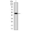
Fig1: Western blot analysis of TrkA on SHSY5Y (1) and SHG-44 (2) cell lysates using anti-TrkA antibody at 1/200 dilution.
Fig2: ICC staining TrkA in PC-12 cells (green). The nuclear counter stain is DAPI (blue). Cells were fixed in paraformaldehyde, permeabilised with 0.25% Triton X100/PBS.
Fig3: Immunohistochemical analysis of paraffin-embedded human liver cancer tissue using anti-TrkA antibody. Counter stained with hematoxylin.
Fig4: Immunohistochemical analysis of paraffin-embedded human stomach cancer tissue using anti-TrkA antibody. Counter stained with hematoxylin.
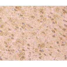
Fig5: Immunohistochemical analysis of paraffin-embedded rat brain tissue using anti-TrkA antibody. Counter stained with hematoxylin.
Fig6: Immunohistochemical analysis of paraffin-embedded mouse brain tissue using anti-TrkA antibody. Counter stained with hematoxylin.
Fig7: Flow cytometric analysis of SHSY5Y cells with TrkA 1/2 antibody at 1/100 dilution (blue) compared with an unlabelled control (cells without incubation with primary antibody; red). Goat anti rabbit IgG (FITC) was used as the secondary antibody.
特别提示:本公司的所有产品仅可用于科研实验,严禁用于临床医疗及其他非科研用途!