Anti-LRRK2 antibody
-
概述
- 产品描述Parkinson's disease is a disorder of movement, cognition and emotion. It is characterized pathologically by neuronal degeneration with Lewy bodies, which are cytoplasmic inclusion bodies containing deposits of aggregated proteins. Mutations in the leucine-rich repeat kinase 2 gene (LRRK2) cause autosomal-dominant parkinsonism, with clinical features of Parkinson's disease and with pleomorphic pathology including deposits of aggregated protein. The LRRK2 protein consists of multiple domains and belongs to the Roco family, a novel group of the Ras/GTPase superfamily. Besides the GTPase (Roc) domain, it contains a predicted kinase domain, with homology to MAP kinase kinase kinases. LRRK2 is localized in the cytoplasm and is associated with cellular membrane structures. The purified LRRK2 protein demonstrates autokinase activity.
- 产品名称Anti-LRRK2 antibody
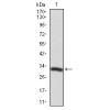
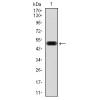
- 分子量286 kDa
- 种属反应性Human, Mouse
- 验证应用WB,ICC,IHC-P
- 抗体类型兔多抗
- 免疫原Synthetic peptide within human LRRK2 aa 2450-2527.
- 偶联Non-conjugated
-
性能
- 形态Liquid
- 浓度1 mg/mL.
- 存放说明Store at +4℃ after thawing. Aliquot store at -20℃ or -80℃. Avoid repeated freeze / thaw cycles.
- 存储缓冲液1*PBS (pH7.4), 0.2% BSA, 50% Glycerol. Preservative: 0.05% Sodium Azide.
- 亚型IgG
- 纯化方式Peptide affinity purified.
- 亚细胞定位Mitochondrion. Lysosome. Golgi apparatus.
- 其它名称augmented in rheumatoid arthritis 17 antibody
AURA17 antibody
Dardarin antibody
Leucine rich repeat kinase 2 antibody
leucine rich repeat serine threonine protein kinase 2 antibody
Leucine-rich repeat serine/threonine-protein kinase 2 antibody
LRRK 2 antibody
LRRK2 antibody
LRRK2_HUMAN antibody
PARK 8 antibody
PARK8 antibody
RIPK7 antibody
ROCO 2 antibody
ROCO2 antibody
more
-
应用
WB: 1:500-1:1,000
ICC: 1:50-1:200
IHC-P: 1:50-1:200
-
Fig1: ICC staining of LRRK2 in A549 cells (green). Formalin fixed cells were permeabilized with 0.1% Triton X-100 in TBS for 10 minutes at room temperature and blocked with 1% Blocker BSA for 15 minutes at room temperature. Cells were probed with the primary antibodyfor 1 hour at room temperature, washed with PBS. Alexa Fluor®488 Goat anti-Rabbit IgG was used as the secondary antibody at 1/1,000 dilution. The nuclear counter stain is DAPI (blue).
Fig2: ICC staining of LRRK2 in N2A cells (green). Formalin fixed cells were permeabilized with 0.1% Triton X-100 in TBS for 10 minutes at room temperature and blocked with 1% Blocker BSA for 15 minutes at room temperature. Cells were probed with the primary antibody for 1 hour at room temperature, washed with PBS. Alexa Fluor®488 Goat anti-Rabbit IgG was used as the secondary antibody at 1/1,000 dilution. The nuclear counter stain is DAPI (blue).
Fig3: ICC staining of LRRK2 in SHSY5Y cells (green). Formalin fixed cells were permeabilized with 0.1% Triton X-100 in TBS for 10 minutes at room temperature and blocked with 1% Blocker BSA for 15 minutes at room temperature. Cells were probed with the primary antibody for 1 hour at room temperature, washed with PBS. Alexa Fluor®488 Goat anti-Rabbit IgG was used as the secondary antibody at 1/1,000 dilution. The nuclear counter stain is DAPI (blue).
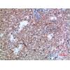
Fig4: Immunohistochemical analysis of paraffin-embedded human liver tissue using anti-LRRK2 antibody. The section was pre-treated using heat mediated antigen retrieval with Tris-EDTA buffer (pH 8.0-8.4) for 20 minutes.The tissues were blocked in 5% BSA for 30 minutes at room temperature, washed with ddH2O and PBS, and then probed with the primary antibody for 30 minutes at room temperature. The detection was performed using an HRP conjugated compact polymer system. DAB was used as the chromogen. Tissues were counterstained with hematoxylin and mounted with DPX.
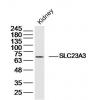
Fig5: Immunohistochemical analysis of paraffin-embedded human kidney tissue using anti-LRRK2 antibody. The section was pre-treated using heat mediated antigen retrieval with Tris-EDTA buffer (pH 8.0-8.4) for 20 minutes.The tissues were blocked in 5% BSA for 30 minutes at room temperature, washed with ddH2O and PBS, and then probed with the primary antibody for 30 minutes at room temperature. The detection was performed using an HRP conjugated compact polymer system. DAB was used as the chromogen. Tissues were counterstained with hematoxylin and mounted with DPX.
Fig6: Immunohistochemical analysis of paraffin-embedded mouse brain tissue using anti-LRRK2 antibody. The section was pre-treated using heat mediated antigen retrieval with Tris-EDTA buffer (pH 8.0-8.4) for 20 minutes.The tissues were blocked in 5% BSA for 30 minutes at room temperature, washed with ddH2O and PBS, and then probed with the primary antibodyfor 30 minutes at room temperature. The detection was performed using an HRP conjugated compact polymer system. DAB was used as the chromogen. Tissues were counterstained with hematoxylin and mounted with DPX.
Fig7: Immunohistochemical analysis of paraffin-embedded mouse cerebellum tissue using anti-LRRK2 antibody. The section was pre-treated using heat mediated antigen retrieval with Tris-EDTA buffer (pH 8.0-8.4) for 20 minutes.The tissues were blocked in 5% BSA for 30 minutes at room temperature, washed with ddH2O and PBS, and then probed with the primary antibody for 30 minutes at room temperature. The detection was performed using an HRP conjugated compact polymer system. DAB was used as the chromogen. Tissues were counterstained with hematoxylin and mounted with DPX.
特别提示:本公司的所有产品仅可用于科研实验,严禁用于临床医疗及其他非科研用途!