Anti-CD99 antibody
-
概述
- 产品描述MIC2, also designated CD99, is a T cell surface protein that is involved in the aggregation of lymphocytes. Two forms of MIC2, which are differentially expressed, are produced by alternative splicing. The major form induces cellular adhesion, whereas the truncated form inhibits the adhesion process. MIC2 regulates the LFA-1/ICAM-1-mediated adhesion of lymphocytes. Overexpression of the truncated form results in downregulated expression of LFA-1. Cells with downregulated MIC2 exhibit a Hodgkin's and Reed-Sternberg (H-RS) phenotype, indicating that MIC2 plays an important role in regulating cell function and morphology.
- 产品名称Anti-CD99 antibody
- 分子量25 kDa (predicted molecular weight: 19 kDa)
- 种属反应性Human,Mouse,Rat
- 验证应用WB,ICC,IHC-P,FC
- 抗体类型兔多抗
- 免疫原Recombinant protein within Human CD99 aa 1-200.
- 偶联Non-conjugated
-
性能
- 形态Liquid
- 浓度1 mg/mL.
- 存放说明Store at +4℃ after thawing. Aliquot store at -20℃. Avoid repeated freeze / thaw cycles.
- 存储缓冲液1*PBS (pH7.4), 0.2% BSA, 50% Glycerol. Preservative: 0.05% Sodium Azide.
- 亚型IgG
- 纯化方式Protein affinity purified.
- 亚细胞定位Membrane.
- 其它名称12E7 antibody
Antigen identified by monoclonal 12E7, Y homolog antibody
Antigen identified by monoclonal antibodies 12E7, F21 and O13 antibody
CD99 antibody
CD99 antigen antibody
CD99 molecule antibody
CD99_HUMAN antibody
Cell surface antigen 12E7 antibody
Cell surface antigen HBA 71 antibody
Cell surface antigen O13 antibody
E2 antigen antibody
HBA71 antibody
MIC 2X antibody
MIC 2Y antibody
MIC2 (monoclonal antibody 12E7) antibody
MIC2 antibody
MIC2X antibody
MIC2Y antibody
MSK5X antibody
Protein MIC2 antibody
Surface antigen MIC2 antibody
T cell surface glycoprotein E2 antibody
T-cell surface glycoprotein E2 antibody
more
-
应用
WB:1:500-1:2,000
ICC:1:200
IHC-P:1:50-1:200
FC:1:50-1:100
-
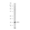
Fig1: Western blot analysis of CD99 on different lysates. Proteins were transferred to a PVDF membrane and blocked with 5% BSA in PBS for 1 hour at room temperature. The primary antibody was used at a 1:500 dilution in 5% BSA at room temperature for 2 hours. Goat Anti-Rabbit IgG - HRP Secondary Antibody (HA1001) at 1:5,000 dilution was used for 1 hour at room temperature.
Positive control:
Lane 1: SiHa cell lysate
Lane 2: HepG2 cell lysate
Fig2: ICC staining CD99 in A549 cells (green). Formalin fixed cells were permeabilized with 0.1% Triton X-100 in TBS for 10 minutes at room temperature and blocked with 1% Blocker BSA for 15 minutes at room temperature. Cells were probed with the antibodyat a dilution of 1:100 for 1 hour at room temperature, washed with PBS. Alexa Fluorc™ 488 Goat anti-Rabbit IgG was used as the secondary antibody at 1/100 dilution. The nuclear counter stain is DAPI (blue).
Fig3: ICC staining CD99 in LOVO cells (green). Formalin fixed cells were permeabilized with 0.1% Triton X-100 in TBS for 10 minutes at room temperature and blocked with 1% Blocker BSA for 15 minutes at room temperature. Cells were probed with the antibodat a dilution of 1:200 for 1 hour at room temperature, washed with PBS. Alexa Fluorc™ 488 Goat anti-Rabbit IgG was used as the secondary antibody at 1/100 dilution. The nuclear counter stain is DAPI (blue).
Fig4: Immunohistochemical analysis of paraffin-embedded rat kidney tissue using anti-CD99 antibody. The section was pre-treated using heat mediated antigen retrieval with Tris-EDTA buffer (pH 8.0-8.4) for 20 minutes.The tissues were blocked in 5% BSA for 30 minutes at room temperature, washed with ddH2O and PBS, and then probed with the antibodyat 1/50 dilution, for 30 minutes at room temperature and detected using an HRP conjugated compact polymer system. DAB was used as the Chromogen. Counter stained with hematoxylin and mounted with DPX.
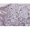
Fig5: Immunohistochemical analysis of paraffin-embedded human esophagus tissue using anti-CD99 antibody. The section was pre-treated using heat mediated antigen retrieval with Tris-EDTA buffer (pH 8.0-8.4) for 20 minutes.The tissues were blocked in 5% BSA for 30 minutes at room temperature, washed with ddH2O and PBS, and then probed with the antibody at 1/200 dilution, for 30 minutes at room temperature and detected using an HRP conjugated compact polymer system. DAB was used as the Chromogen. Counter stained with hematoxylin and mounted with DPX.
Fig6: Immunohistochemical analysis of paraffin-embedded human pancreas tissue using anti-CD99 antibody. The section was pre-treated using heat mediated antigen retrieval with Tris-EDTA buffer (pH 8.0-8.4) for 20 minutes.The tissues were blocked in 5% BSA for 30 minutes at room temperature, washed with ddH2O and PBS, and then probed with the antibodyat 1/200 dilution, for 30 minutes at room temperature and detected using an HRP conjugated compact polymer system. DAB was used as the Chromogen. Counter stained with hematoxylin and mounted with DPX.
Fig7: Immunohistochemical analysis of paraffin-embedded mouse testis tissue using anti-CD99 antibody. The section was pre-treated using heat mediated antigen retrieval with Tris-EDTA buffer (pH 8.0-8.4) for 20 minutes.The tissues were blocked in 5% BSA fo
Fig8: Flow cytometric analysis of CD99 was done on 293T cells. The cells were fixed, permeabilized and stained with CD99 antibody at 1/100 dilution (red) compared with an unlabelled control (cells without incubation with primary antibody; black). After in
特别提示:本公司的所有产品仅可用于科研实验,严禁用于临床医疗及其他非科研用途!