Anti-Neurabin 2 antibody
-
概述
- 产品描述Seems to act as a scaffold protein in multiple signaling pathways. Modulates excitatory synaptic transmission and dendritic spine morphology. Binds to actin filaments (F-actin) and shows cross-linking activity. Binds along the sides of the F-actin. May play an important role in linking the actin cytoskeleton to the plasma membrane at the synaptic junction. Believed to target protein phosphatase 1/PP1 to dendritic spines, which are rich in F-actin, and regulates its specificity toward ion channels and other substrates, such as AMPA-type and NMDA-type glutamate receptors. Plays a role in regulation of G-protein coupled receptor signaling, including dopamine D2 receptors and alpha-adrenergic receptors. May establish a signaling complex for dopaminergic neurotransmission through D2 receptors by linking receptors downstream signaling molecules and the actin cytoskeleton. Binds to ADRA1B and RGS2 and mediates regulation of ADRA1B signaling. May confer to Rac signaling specificity by binding to both, RacGEFs and Rac effector proteins. Probably regulates p70 S6 kinase activity by forming a complex with TIAM1 (By similarity). Required for hepatocyte growth
- 产品名称Anti-Neurabin 2 antibody
- 分子量89 KDa
- 种属反应性Human,Mouse,Rat,Dog,Pig,Sheep
- 验证应用WB,IHC-P
- 抗体类型兔多抗
- 免疫原KLH conjugated synthetic peptide derived from human Spinophilin/Neurabin 2 358-460/815
- 偶联Non-conjugated
-
性能
- 形态Liquid
- 浓度1 mg/mL.
- 存放说明Store at -20℃ for one year. Avoid repeated freeze/thaw cycles. The lyophilized antibody is stable at room temperature for at least one month and for greater than a year when kept at -20℃. When reconstituted in sterile pH 7.4 0.01M PBS or diluent of antibody the antibody is stable for at least two weeks at 2-4℃.
- 存储缓冲液0.01M TBS(pH7.4) with 1% BSA, 0.03% Proclin300 and 50% Glycerol.
- 亚型IgG
- 纯化方式affinity purified by Protein A
- 亚细胞定位Cytoplasm, cytoskeleton (By similarity). Nucleus (By similarity). Cell projection, dendritic spine (By similarity). Cell junction, synapse. Cell junction, adherens junction (By similarity). Cytoplasm. Cell membrane. Cell projection, lamellipodium. Cell pr
- 其它名称FLJ30345
NEB2_HUMAN
Neurabin II
Neurabin-2
Neurabin-II
Neurabin2
NeurabinII
Neurabin II
Neural tissue specific F actin binding protein II
p130
PP1bp134
PPP1R6
PPP1R9
PPP1R9B
Protein phosphatase 1 regulatory subunit 9B
SPINO
Spinophilin.
more
-
应用
WB:1:500-2000
IHC-P:1:400-800
-
Fig1: Paraformaldehyde-fixed, paraffin embedded (mouse brain tissue); Antigen retrieval by boiling in sodium citrate buffer (pH6.0) for 15min; Block endogenous peroxidase by 3% hydrogen peroxide for 20 minutes; Blocking buffer (normal goat serum) at 37℃ for 30min; Antibody incubation with (Neurabin 2) Polyclonal Antibody, Unconjugatedat 1:400 overnight at 4℃, followed by operating according to SP Kit(Rabbit) (sp-0023) instructionsand DAB staining.
Fig2: Tissue/cell: rat brain tissue; 4% Paraformaldehyde-fixed and paraffin-embedded;
Antigen retrieval: citrate buffer ( 0.01M, pH 6.0 ), Boiling bathing for 15min; Block endogenous peroxidase by 3% Hydrogen peroxide for 30min; Blocking buffer (normal goat serum,C-0005) at 37℃ for 20 min;
Incubation: Anti-Neurabin 2 Polyclonal Antibody, Unconjugated1:200, overnight at 4℃, followed by conjugation to the secondary antibody(SP-0023) and DAB(C-0010) staining
Fig3: Paraformaldehyde-fixed, paraffin embedded (rat brain tissue); Antigen retrieval by boiling in sodium citrate buffer (pH6.0) for 15min; Block endogenous peroxidase by 3% hydrogen peroxide for 20 minutes; Blocking buffer (normal goat serum) at 37℃ for 30min; Antibody incubation with (Neurabin 2) Polyclonal Antibody, Unconjugated at 1:400 overnight at 4℃, followed by operating according to SP Kit(Rabbit) (sp-0023) instructionsand DAB staining.
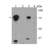
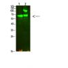
Fig4: Sample:
Lung (Mouse) Lysate at 40 ug
Primary: Anti- Neurabin 2 at 1/1000 dilution
Secondary: IRDye800CW Goat Anti-Rabbit IgG at 1/20000 dilution
Predicted band size: 89 kD
Observed band size: 89 kD
特别提示:本公司的所有产品仅可用于科研实验,严禁用于临床医疗及其他非科研用途!


