Anti-AKT1S1 antibody [6H1C6]
-
概述
- 产品描述Subunit of mTORC1, which regulates cell growth and survival in response to nutrient and hormonal signals. mTORC1 is activated in response to growth factors or amino acids. Growth factor-stimulated mTORC1 activation involves a AKT1-mediated phosphorylation of TSC1-TSC2, which leads to the activation of the RHEB GTPase that potently activates the protein kinase activity of mTORC1. Amino acid-signaling to mTORC1 requires its relocalization to the lysosomes mediated by the Ragulator complex and the Rag GTPases. Activated mTORC1 up-regulates protein synthesis by phosphorylating key regulators of mRNA translation and ribosome synthesis. mTORC1 phosphorylates EIF4EBP1 and releases it from inhibiting the elongation initiation factor 4E (eiF4E). mTORC1 phosphorylates and activates S6K1 at 'Thr-389', which then promotes protein synthesis by phosphorylating PDCD4 and targeting it for degradation. Within mTORC1, AKT1S1 negatively regulates mTOR activity in a manner that is dependent on its phosphorylation state and binding to 14-3-3 proteins. Inhibits RHEB-GTP-dependent mTORC1 activation. Substrate for AKT1 phosphorylation, but can also be activated by AKT1-independent mechanisms. May also play a role in nerve growth factor-mediated neuroprotection.
- 产品名称Anti-AKT1S1 antibody [6H1C6]
- 分子量27.4kDa
-
-
种属反应性Human
-
验证应用WB,IHC-P,ICC
-
抗体类型小鼠单抗
-
免疫原Purified recombinant fragment of human AKT1S1 (AA: 92-276) expressed in E. Coli.
-
偶联Non-conjugated
-
Anti-AKT1S1 antibody性能
-
形态Liquid
-
浓度1 mg/mL
-
存放说明Store at +4℃ after thawing. Aliquot store at -20℃. Avoid repeated freeze / thaw cycles.
-
存储缓冲液1*PBS with 0.05% sodium azide.
-
亚型IgG1
-
纯化方式Protein G purified.
-
亚细胞定位Cytoplasm.
-
-
其它名称
more
-
应用
WB: 1:500-1:2,000
IHC-P: 1:50-1:200
ICC: 1:50-1:200
-
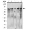
Fig1: Western blot analysis of AKT1S1 against human AKT1S1 (AA: 92-276) recombinant protein. Proteins were transferred to a PVDF membrane and blocked with 5% BSA in PBS for 1 hour at room temperature. The primary antibody was used in 5% BSA at room temperature for 2 hours. Goat Anti-Mouse IgG - HRP Secondary Antibody at 1:5,000 dilution was used for 1 hour at room temperature.
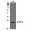
Fig2: Western blot analysis of AKT1S1 against HEK293 (1) and AKT1S1 (AA: 92-276)-hIgGFc transfected HEK293 (2) cell lysate. Proteins were transferred to a PVDF membrane and blocked with 5% BSA in PBS for 1 hour at room temperature. The primary antibody was used in 5% BSA at room temperature for 2 hours. Goat Anti-Mouse IgG - HRP Secondary Antibody at 1:5,000 dilution was used for 1 hour at room temperature.
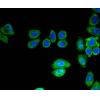
Fig3: Immunocytochemistry staining of AKT1S1 in Hela cells (green). Formalin fixed cells were permeabilized with 0.1% Triton X-100 in TBS for 10 minutes at room temperature and blocked with 1% Blocker BSA for 15 minutes at room temperature. Cells were probed with the primary antibody for 1 hour at room temperature, washed with PBS. Alexa Fluor®488 Goat anti-Mouse IgG was used as the secondary antibody at 1/1,000 dilution. The nuclear counter stain is DAPI (blue), Actin filaments have been labeled with Alexa Fluor- 555 phalloidin (red).
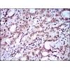
Fig4: Immunohistochemical analysis of paraffin-embedded colon cancer tissue using anti-AKT1S1 antibody. The section was pre-treated using heat mediated antigen retrieval with Tris-EDTA buffer (pH 8.0) for 20 minutes. The tissues were blocked in 5% BSA for 30 minutes at room temperature, washed with ddH2O and PBS, and then probed with the primary antibody for 30 minutes at room temperature. The detection was performed using an HRP conjugated compact polymer system. DAB was used as the chromogen. Tissues were counterstained with hematoxylin and mounted with DPX.
Fig5: Immunohistochemical analysis of paraffin-embedded endometrial cancer tissue using anti-AKT1S1 antibody. The section was pre-treated using heat mediated antigen retrieval with Tris-EDTA buffer (pH 8.0) for 20 minutes. The tissues were blocked in 5% BSA for 30 minutes at room temperature, washed with ddH2O and PBS, and then probed with the primary antibody for 30 minutes at room temperature. The detection was performed using an HRP conjugated compact polymer system. DAB was used as the chromogen. Tissues were counterstained with hematoxylin and mounted with DPX.
特别提示:本公司的所有产品仅可用于科研实验,严禁用于临床医疗及其他非科研用途!

![Anti-AKT1S1 antibody [6H1C6]](images/202012/goods_img/92818_G_1608035137100.jpg)