Anti-SIRT3 antibody
-
概述
- 产品描述The Silent Information Regulator (SIR2) family of genes are highly conserved from prokaryotes to eukaryotes and are involved in diverse processes, including transcriptional regulation, cell cycle progression, DNA-damage repair and aging. In S. cerevisiae, Sir2p deacetylates histones in an NAD-dependent manner, which regulates silencing at the telomeric, rDNA and silent mating-type loci. Sir2p is the founding member of a large family, designated sirtuins, which contain a conserved catalytic domain. The human homologues, which include SIRT1-7, are divided into four main branches: SIRT1-3 are class I, SIRT4 is class II, SIRT5 is class III and SIRT6-7 are class IV. SIRT3 is a NAD-dependent deacetylase that contains one deacetylase sirtuin-type domain. The SIRT3 protein is widely expressed and localizes to the mitochondira where it is processed by mitochondrial processing peptidase (MPP) to yield a final product. This processing is most-likely necessary for its enzymatic activity.
- 产品名称Anti-SIRT3 antibody
- 分子量44/28kDa
- 种属反应性Human,Mouse
- 验证应用WB,ICC,IHC-P,FC
- 抗体类型兔多抗
- 免疫原peptide
- 偶联Non-conjugated
-
性能
- 形态Liquid
- 浓度1 mg/mL.
- 存放说明Store at +4℃ after thawing. Aliquot store at -20℃ or -80℃. Avoid repeated freeze / thaw cycles.
- 存储缓冲液1*PBS (pH7.4), 0.2% BSA, 40% Glycerol. Preservative: 0.05% Sodium Azide.
- 亚型IgG
- 纯化方式Peptide affinity purified
- 亚细胞定位Mitochondrion matrix
- 其它名称
- hSIRT 3 antibody
- hSIRT3 antibody
- Mitochondrial nicotinamide adenine dinucleotide dependent deacetylase antibody
more
-
应用
WB: 1:1,000
ICC: 1:50-1:200
IHC-P: 1:50-1:200
FC: 1:50-1:100
-
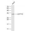
Fig1: Western blot analysis on different lysates using anti-SIRT3 rabbit polyclonal antibody. Positive control:
Lane 1: A172
Lane 2: Mouse liver
Lane 3: NIH/3T3
Lane 4: Mouse kidney
Lane 5: F9
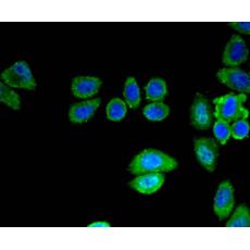
Fig2: Immunocytochemical staining of MCF-7 cells using anti-SIRT3 rabbit polyclonal antibody.
Fig3: Immunocytochemical staining of HepG2 cells using anti-SIRT3 rabbit polyclonal antibody.
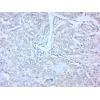
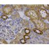
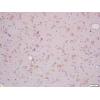
Fig4: Immunohistochemical analysis of paraffin- embedded human kidney tissue using anti-SIRT3 rabbit polyclonal antibody.
Fig5: Immunohistochemical analysis of paraffin- embedded mouse kidney tissue using anti-SIRT3 rabbit polyclonal antibody.
Fig6: Flow cytometric analysis of HepG2 cells with SIRT3 antibody at 1/50 dilution (blue) compared with an unlabelled control (cells without incubation with primary antibody; red). Alexa Fluor 488-conjugated Goat anti rabbit IgG was used as the secondary antibody.
特别提示:本公司的所有产品仅可用于科研实验,严禁用于临床医疗及其他非科研用途!


