Anti-TIP60 antibody [H7-A11]
-
概述
- 产品描述MOZ (monocytic leukemia zinc finger protein) is a chromatin-associated histone acetyltransferase (HAT) that regulates chromatin remodeling and transcription. The MOZ gene was initially isolated as a consequence of two variant translocations that were identified in a distinct subtype of acute myeloid leukemias and resulted in the formation of MOZ fusion proteins. These fusions involve the HAT domain of MOZ with the activation domain of either transcriptional coactivator protein TIF2/GRIP1 or CBP, and lead to enhanced transcriptional activation by a mechanism involving aberrant histone acetylation. Additional MOZ related proteins, including MORF (MOZ related factor) and TIP60 (TAT interacting proteins 60), share significant similarities with MOZ including the putuative HAT domain. MORF also contains a strong transcriptional repression domain at its N terminus and a highly potent activation domain at the C terminus, suggesting that MORF has both HAT activity and contributes to the regulation of transcriptional activation. TIP60 was originally identified as a coactivator for the HIV TAT protein and also functions as a nuclear hormone receptor coactivator that enhances ligand dependent steroid receptor-mediated transactivation involving the androgen, estrogen and progesterone receptors.
- 产品名称Anti-TIP60 antibody [H7-A11]
- 分子量59 kDa
- 种属反应性Human
- 验证应用WB,ICC,FC
- 抗体类型小鼠单抗
- 免疫原Recombinant protein
- 偶联Non-conjugated
-
性能
- 形态Liquid
- 浓度2 mg/mL.
- 存放说明Store at +4℃ after thawing. Aliquot store at -20℃ or -80℃. Avoid repeated freeze / thaw cycles.
- 存储缓冲液1*TBS (pH7.4), 1%BSA, 40%Glycerol. Preservative: 0.05% Sodium Azide.
- 亚型IgG1
- 纯化方式Protein A purified.
- 亚细胞定位Nucleus, Cytoplasm
- 其它名称
- 60 kDa Tat interactive protein antibody
- 60 kDa Tat-interactive protein antibody
- cPLA(2) interacting protein antibody
more
-
应用
WB: 1:500-1:2,000
ICC: 1:50-1:200
FC: 1:50-1:100
-
Fig1: Western blot analysis of TIP60 on human TIP60 recombinant protein using anti-TIP60 antibody at 1/1,000 dilution.
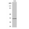
Fig2: Western blot analysis of TIP60 on HEK293 (1) and TIP60-hIgGFc transfected HEK293 (2) cell lysate using anti-TIP60 antibody at 1/1,000 dilution.
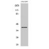
Fig3: Western blot analysis of TIP60 on Hela cell lysate using anti-TIP60 antibody at 1/1,000 dilution.
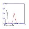
Fig4: Flow cytometric analysis of Hela cells with TIP60 antibody at 1/100 dilution (green) compared with an unlabelled control (cells without incubation with primary antibody; red).
特别提示:本公司的所有产品仅可用于科研实验,严禁用于临床医疗及其他非科研用途!

![Anti-TIP60 antibody [H7-A11]](images/202012/goods_img/92754_G_1607942055129.jpg)