Anti-Tyrosinase antibody [C2-B5]
-
概述
- 产品描述This is a copper-containing oxidase that functions in the formation of pigments such as melanins and other polyphenolic compounds. Catalyzes the initial and rate limiting step in the cascade of reactions leading to melanin production from tyrosine. In addition to hydroxylating tyrosine to DOPA (3,4-dihydroxyphenylalanine), also catalyzes the oxidation of DOPA to DOPA-quinone, and possibly the oxidation of DHI (5,6-dihydroxyindole) to indole-5,6 quinone. Studies have shown tyrosinase to be a very specific marker for melanomas, not cross reacting with any other tumors or normal tissues tested Other studies have shown tyrosinase to be a more sensitive marker when compared to HMB-45 and MART-1. It has also shown to label a higher percentage of desmoplastic melanomas than HMB-45. However, both tyrosinase and MART-1 negative staining was seen in those variants without an epidermal component. Unlike HMB-45, neither tyrosinase or MART-1 discriminates between activated or resting melanocytes. In conclusion, tyrosinase appears to be a superior melanoma marker when compared to HMB-45.
- 产品名称Anti-Tyrosinase antibody [C2-B5]
- 分子量60 kDa
- 种属反应性Human,Mouse
- 验证应用WB,ICC,IHC-P,FC
- 抗体类型小鼠单抗
- 免疫原Peptide linked to KLH
- 偶联Non-conjugated
-
性能
- 形态Liquid
- 浓度2 mg/mL.
- 存放说明Store at +4℃ after thawing. Aliquot store at -20℃ or -391℃. Avoid repeated freeze / thaw cycles.
- 存储缓冲液1*PBS (pH7.4), 0.2% BSA, 40% Glycerol. Preservative: 0.05% Sodium Azide.
- 亚型IgG1
- 纯化方式Protein A purified.
- 亚细胞定位Membrane.
- 其它名称
- ATN antibody
- CMM8 antibody
- LB24 AB antibody
more
-
应用
WB: 1:1,000-1:2,000
ICC: 1:50-1:200
IHC-P: 1:50-1:200
FC: 1:50-1:100
-
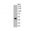
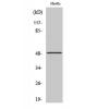
Fig1: Western blot analysis of tyrosinase on melanoma cells lysates using anti- tyrosinase antibody at 1/1,000 dilution.
Fig2: ICC staining tyrosinase in B16F1 cells (red). The nuclear counter stain is DAPI (blue). Cells were fixed in paraformaldehyde, permeabilised with 0.25% Triton X100/PBS.
Fig3: Immunohistochemical analysis of paraffin-embedded human tyrosinase tissue using anti- tyrosinase antibody. Counter stained with hematoxylin.
Fig4: Flow cytometric analysis of B16F1 cells with tyrosinase antibody at 1/100 dilution (red) compared with an unlabelled control (cells without incubation with primary antibody; black). Alexa Fluor 488-conjugated Goat anti mouse IgG was used as the secondary antibody
特别提示:本公司的所有产品仅可用于科研实验,严禁用于临床医疗及其他非科研用途!


![Anti-Tyrosinase antibody [C2-B5]](images/202012/goods_img/92463_G_1607331156568.jpg)


