Anti-TGM6 antibody
-
概述
- 产品描述Transglutaminase 6 is a protein that in humans is encoded by the TGM6 gene. The protein encoded by this gene belongs to the transglutaminase superfamily. It catalyzes the cross-linking of proteins and the conjugation of polyamines to proteins. Mutations in this gene are associated with spinocerebellar ataxia type 35 (SCA35). Alternatively spliced transcript variants encoding different isoforms have been found for this gene.
- 产品名称Anti-TGM6 antibody
- 分子量79/71 kDa
- 种属反应性Human,Mouse,Rat
- 验证应用WB,ICC,IHC-P,FC
- 抗体类型兔多抗
- 免疫原Peptide
- 偶联Non-conjugated
-
性能
- 形态Liquid
- 浓度1 mg/mL.
- 存放说明Store at +4℃ after thawing. Aliquot store at -20℃ or -80℃. Avoid repeated freeze / thaw cycles.
- 存储缓冲液1*PBS (pH7.4), 0.2% BSA, 50% Glycerol. Preservative: 0.05% Sodium Azide.
- 亚型IgG
- 纯化方式Peptide affinity purified
- 亚细胞定位Cytoplasm.
- 其它名称Protein-glutamine gamma-glutamyltransferase 6 antibody
TG6 antibody
TGase Y antibody
TGase-3-like antibody
TGase-6 antibody
TGM3L_HUMAN antibody
TGM6 antibody
TGY antibody
Transglutaminase Y antibody
Transglutaminase-3-like antibody
Transglutaminase-6 antibody
more
-
应用
WB:1:500
ICC: 1:50-1:200
IHC-P: 1:50-1:200
FC: 1:50-1:100
-
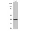
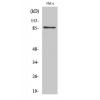
Fig1: Western blot analysis of TGM6 on different tissue lysates using anti-TGM6 antibody at 1/100 dilution.
Positive control:
Lane 1 : Mouse brain
Lane 2 : Mouse brain
Lane 3 : Mouse spleen
Lane 4 : Mouse spleen
Lane 5 : Mouse testis
Fig2: ICC staining TGM6 in A549 cells (green). The nuclear counter stain is DAPI (blue). Cells were fixed in paraformaldehyde, permeabilised with 0.25% Triton X100/PBS.
Fig3: ICC staining TGM6 in SH-SY5Y cells (green). The nuclear counter stain is DAPI (blue). Cells were fixed in paraformaldehyde, permeabilised with 0.25% Triton X100/PBS.
Fig4: ICC staining TGM6 in 293T cells (green). The nuclear counter stain is DAPI (blue). Cells were fixed in paraformaldehyde, permeabilised with 0.25% Triton X100/PBS.
Fig5: Immunohistochemical analysis of paraffin-embedded human kidney tissue using anti-TGM6 antibody. Counter stained with hematoxylin.
Fig6: Immunohistochemical analysis of paraffin-embedded mouse testis tissue using anti-TGM6 antibody. Counter stained with hematoxylin.
Fig7: Immunohistochemical analysis of paraffin-embedded mouse brain tissue using anti-TGM6 antibody. Counter stained with hematoxylin.
Fig8: Immunohistochemical analysis of paraffin-embedded rat brain tissue using anti-TGM6 antibody. Counter stained with hematoxylin.
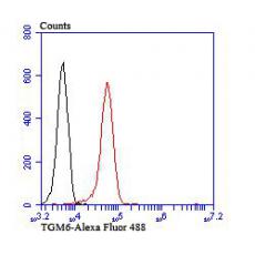
Fig9: Flow cytometric analysis of 293T cells with TGM6 antibody at 1/100 dilution (red) compared with an unlabelled control (cells without incubation with primary antibody; black). Alexa Fluor 488-conjugated Goat anti rabbit IgG was used as the secondary
特别提示:本公司的所有产品仅可用于科研实验,严禁用于临床医疗及其他非科研用途!