Anti-ATP2A1 antibody [H6-D1]
-
概述
- 产品描述ATP dependent calcium pumps are responsible, in part, for the maintenance of low cytoplasmic free calcium concentrations. The ATP pumps that reside in intracellular organelles are encoded by a family of structurally related enzymes, termed the sarcoplasmic or endoplasmic reticulum calcium (SERCA) ATPases. The sarcoplasmic reticulum of striated muscle is a specialized intracellular membrane system that plays a critical role in the contraction and relaxation of muscle. The SERCAs mediate Ca2+ uptake into intracellular stores. SERCA-mediated Ca2+ uptake induces and maintains muscular relaxation. The SERCA1 gene is exclusively expressed in type II (fast) skeletal muscle. The SERCA2 gene is subject to tissue-dependent processing which is responsible for the generation of the SERCA2a muscle-specific form expressed in type I (slow) skeletal, cardiac and smooth muscle, and the SERCA2b isoform expressed in all cell types. The SERCA3 gene is not as well characterized and is found in non-muscle cells. SERCA2 plays an important part in regulating cardiac contractile function. SERCA3 is an isoform expressed in several cell types including platelets, lymphoid cells and mast cells. SERCA1, SERCA2 and SERCA3 all undergo alternative splicing.
- 产品名称Anti-ATP2A1 antibody [H6-D1]
- 分子量110 kDa
- 种属反应性Human,Mouse,Monkey
- 验证应用WB,FC
- 抗体类型小鼠单抗
- 免疫原Recombinant protein
- 偶联Non-conjugated
-
性能
- 形态Liquid
- 浓度2 mg/mL.
- 存放说明Store at +4℃ after thawing. Aliquot store at -20℃ or -80℃. Avoid repeated freeze / thaw cycles.
- 存储缓冲液1*TBS (pH7.4), 1%BSA, 40%Glycerol. Preservative: 0.05% Sodium Azide.
- 亚型IgG1
- 纯化方式Protein A purified.
- 亚细胞定位Endoplasmic reticulum membrane. Sarcoplasmic reticulum membrane.
- 其它名称
- fast twitch skeletal muscle isoform antibody
- AT2A1_HUMAN antibody
- ATP2A antibody
more
-
应用
WB: 1:500-1:2,000
FC: 1:100-1:200
-
Fig1: Western blot analysis of ATP2A1 on human ATP2A1 recombinant protein using anti-ATP2A1 antibody at 1/1,000 dilution.
Fig2: Western blot analysis of ATP2A1 on HEK293 (1) and ATP2A1-hIgGFc transfected HEK293 (2) cell lysate using anti-ATP2A1 antibody at 1/1,000 dilution.
Fig3: Western blot analysis of ATP2A1 on different cell lysate using anti-ATP2A1 antibody at 1/1,000 dilution.
Positive control:
Lane 1: C2C12
Lane 2: COS7
Lane 3: Hela
Lane 4: K562
Lane 5: Jurkat
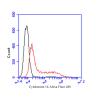
Fig4: Flow cytometric analysis of Hela cells with ATP2A1 antibody at 1/100 dilution (green) compared with an unlabelled control (cells without incubation with primary antibody; red).
特别提示:本公司的所有产品仅可用于科研实验,严禁用于临床医疗及其他非科研用途!

![Anti-ATP2A1 antibody [H6-D1]](images/202012/goods_img/92133_G_1606985534906.jpg)