Anti-Desmoglein 3 antibody
-
概述
- 产品描述Pemphigus is an autoimmune disease of skin adhesion associated with auto-antibodies against a number of keratinocyte antigens, such as the adhesion molecules desmoglein (dsg) 1 and 3 and acetylcholine receptors. Desmogleins, type I membrane proteins, are important for cell adhesion and are expressed in great abundance at the desmosomes, which are adhesive cell junctions. Desmogleins belong to the cadherin family and consist of dsg1, dsg2 and dsg3. Calcium binds to the putative calcium binding sites at the extracellular N-terminal domain, which has cadherin-like repeats. Unlike normal human keratinocytes, the squamous cell carcinoma cells exhibit diminished or un-usual expression of dsg3 and dsg1, which bear pemphigus vulgaris and pemphigus foliaceus antigens, respectively. Several carcinoma cell lines constantly express dsg2 and dsg3 mRNA, whereas cultured normal human keratinocytes always express dsg1 and dsg3 mRNA, with or without dsg2 mRNA. This expression pattern indicates that desmoglein isoforms exhibit abnormal expression and may be related to tumor cell kinetics, such as cell invasion and metastasis. dsg2 is the fundamental dsg common to all desmosome-possessing tissues and is the largest desmoglein in the family.
- 产品名称Anti-Desmoglein 3 antibody
- 分子量Predicted band size 108 kDa.
- 种属反应性Human, Mouse, Rat
- 验证应用WB,ICC,IHC-P,FC
- 抗体类型兔多抗
- 免疫原Recombinant protein within Human DSG3 aa 700-880.
- 偶联Non-conjugated
-
性能
- 形态Liquid
- 浓度1 mg/mL.
- 存放说明Store at +4℃ after thawing. Aliquot store at -20℃. Avoid repeated freeze / thaw cycles.
- 存储缓冲液1*PBS (pH7.4), 0.2% BSA, 50% Glycerol. Preservative: 0.05% Sodium Azide.
- 亚型IgG
- 纯化方式Protein affinity purified.
- 亚细胞定位Cell membrane.
- 其它名称
- 130 kD pemphigus vulgaris antigen antibody
- 130 kDa pemphigus vulgaris antigen antibody
- Balding antibody
more
-
应用
WB: 1:500-1:2000
ICC:1:100-1:200
IHC-P:1:50-1:200
FC: 1:50-1:100
-
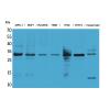
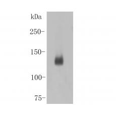
Fig1: Western blot analysis of DSG3 on A431 cell lysates. Proteins were transferred to a PVDF membrane and blocked with 5% BSA in PBS for 1 hour at room temperature. The primary antibody was used in 5% BSA at room temperature for 2 hours. Goat Anti-Rabbit IgG - HRP Secondary Antibody (HA1001) at 1:5,000 dilution was used for 1 hour at room temperature.
Fig2: ICC staining DSG3 in A549 cells (green). Formalin fixed cells were permeabilized with 0.1% Triton X-100 in TBS for 10 minutes at room temperature and blocked with 1% Blocker BSA for 15 minutes at room temperature. Cells were probed with the antibody at a dilution of 1:200 for 1 hour at room temperature, washed with PBS. Alexa Fluorc™ 488 Goat anti-Rabbit IgG was used as the secondary antibody at 1/100 dilution. The nuclear counter stain is DAPI (blue).
Fig3: ICC staining DSG3 in Hela cells (green). Formalin fixed cells were permeabilized with 0.1% Triton X-100 in TBS for 10 minutes at room temperature and blocked with 1% Blocker BSA for 15 minutes at room temperature. Cells were probed with the antibodyat a dilution of 1:200 for 1 hour at room temperature, washed with PBS. Alexa Fluorc™ 488 Goat anti-Rabbit IgG was used as the secondary antibody at 1/100 dilution. The nuclear counter stain is DAPI (blue).
Fig4: Immunohistochemical analysis of paraffin-embedded rat esophagus tissue using anti-DSG3 antibody. The section was pre-treated using heat mediated antigen retrieval with Tris-EDTA buffer (pH 8.0-8.4) for 20 minutes.The tissues were blocked in 5% BSA for 30 minutes at room temperature, washed with ddH2O and PBS, and then probed with the antibody at 1/50 dilution, for 30 minutes at room temperature and detected using an HRP conjugated compact polymer system. DAB was used as the chrogen. Counter stained with hematoxylin and mounted with DPX.
Fig5: Immunohistochemical analysis of paraffin-embedded human esophagus cancer tissue using anti-DSG3 antibody. The section was pre-treated using heat mediated antigen retrieval with Tris-EDTA buffer (pH 8.0-8.4) for 20 minutes.The tissues were blocked in 5% BSA for 30 minutes at room temperature, washed with ddH2O and PBS, and then probed with the antibody at 1/200 dilution, for 30 minutes at room temperature and detected using an HRP conjugated compact polymer system. DAB was used as the chrogen. Counter stained with hematoxylin and mounted with DPX.
Fig6: Immunohistochemical analysis of paraffin-embedded mouse skin tissue using anti-DSG3 antibody. The section was pre-treated using heat mediated antigen retrieval with Tris-EDTA buffer (pH 8.0-8.4) for 20 minutes.The tissues were blocked in 5% BSA for 30 minutes at room temperature, washed with ddH2O and PBS, and then probed with the antibody ( at 1/50 dilution, for 30 minutes at room temperature and detected using an HRP conjugated compact polymer system. DAB was used as the chrogen. Counter stained with hematoxylin and mounted with DPX.
Fig7: Flow cytometric analysis of DSG3 was done on Hela cells. The cells were fixed, permeabilized and stained with the primary antibody(red). After incubation of the primary antibody at room temperature for an hour, the cells were stained with a Alexa Fluor 488-conjugated goat anti-rabbit IgG Secondary antibody at 1/500 dilution for 30 minutes.Unlabelled sample was used as a control (cells without incubation with primary antibody; blue).
特别提示:本公司的所有产品仅可用于科研实验,严禁用于临床医疗及其他非科研用途!