Anti-delta 1 Catenin/CAS antibody
-
概述
- 产品描述This gene encodes a member of the Armadillo protein family, which function in adhesion between cells and signal transduction. Multiple translation initiation codons and alternative splicing result in many different isoforms being translated. Not all of the full-length natures of the described transcript variants have been determined. Read-through transcription also exists between this gene and the neighboring upstream thioredoxin-related transmembrane protein 2 (TMX2) gene. Binds to and inhibits the transcriptional repressor ZBTB33, which may lead to activation of target genes of the Wnt signaling pathway (By similarity). Associates with and regulates the cell adhesion properties of both C-, E- and N-cadherins, being critical for their surface stability. Implicated both in cell transformation by SRC and in ligand-induced receptor signaling through the EGF, PDGF, CSF-1 and ERBB2 receptors. Promotes GLIS2 C-terminal cleavage.
- 产品名称Anti-delta 1 Catenin/CAS antibody
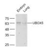
- 分子量Predicted band size 108 kDa.
- 种属反应性Human,Mouse,Rat
- 验证应用WB,ICC,IHC-P,FC
- 抗体类型兔多抗
- 免疫原Synthetic peptide within human delta 1 Catenin/CAS aa 50-120.
- 偶联Non-conjugated
-
性能
- 形态Liquid
- 浓度1 mg/ml.
- 存放说明Store at +4℃ after thawing. Aliquot store at -20℃. Avoid repeated freeze / thaw cycles.
- 存储缓冲液1*PBS (pH7.4), 0.2% BSA, 50% Glycerol. Preservative: 0.05% Sodium Azide.
- 亚型IgG
- 纯化方式Peptide affinity purified.
- 亚细胞定位Cell membrane, Cytoplasm, Membrane, Nucleus.
- 其它名称
- Cadherin associated Src substrate antibody
- Cadherin-associated Src substrate antibody
- CAS antibody
- Catenin (cadherin associated protein) delta 1 antibody
- Catenin delta 1 antibody
- Catenin delta antibody
- Catenin delta-1 antibody
- CTND1_HUMAN antibody
- CTNND 1 antibody
- CTNND antibody
- CTNND1 antibody
- delta 1 Catenin antibody
- KIAA0384 antibody
- p120 antibody
- P120 CAS antibody
- p120 catenin antibody
- P120 CTN antibody
- p120(cas) antibody
- p120(ctn) antibody
- P120CAS antibody
- P120CTN antibody
pick up
-
应用
WB: 1:500-1:2000
ICC: 1:100-1:500
IHC-P: 1:100-1:500
FC: 1:50-1:200
-
Fig1: Western blot analysis of delta 1 Catenin/CAS on HUVEC cell lysate. Proteins were transferred to a PVDF membrane and blocked with 5% BSA in PBS for 1 hour at room temperature. The primary antibody was used in 5% BSA at room temperature for 2 hours. Goat Anti-Rabbit IgG - HRP Secondary Antibody (HA1001) at 1:5,000 dilution was used for 1 hour at room temperature.
Fig2: Western blot analysis of delta 1 Catenin/CAS on mouse stomach tissue lysate. Proteins were transferred to a PVDF membrane and blocked with 5% BSA in PBS for 1 hour at room temperature. The primary antibody was used in 5% BSA at room temperature for 2 hours. Goat Anti-Rabbit IgG - HRP Secondary Antibody (HA1001) at 1:5,000 dilution was used for 1 hour at room temperature.
Fig3: Western blot analysis of delta 1 Catenin/CAS on rat skin tissue lysate. Proteins were transferred to a PVDF membrane and blocked with 5% BSA in PBS for 1 hour at room temperature. The primary antibody was used in 5% BSA at room temperature for 2 hours. Goat Anti-Rabbit IgG - HRP Secondary Antibody (HA1001) at 1:5,000 dilution was used for 1 hour at room temperature.
Fig4: ICC staining of delta 1 Catenin/CAS in A431 cells (green). Formalin fixed cells were permeabilized with 0.1% Triton X-100 in TBS for 10 minutes at room temperature and blocked with 1% Blocker BSA for 15 minutes at room temperature. Cells were probed with the primary antibody for 1 hour at room temperature, washed with PBS. Alexa Fluor®488 Goat anti-Rabbit IgG was used as the secondary antibody at 1/100 dilution. The nuclear counter stain is DAPI (blue).
Fig5: ICC staining of delta 1 Catenin/CAS in Siha cells (green). Formalin fixed cells were permeabilized with 0.1% Triton X-100 in TBS for 10 minutes at room temperature and blocked with 1% Blocker BSA for 15 minutes at room temperature. Cells were probed with the primary antibodyfor 1 hour at room temperature, washed with PBS. Alexa Fluor®488 Goat anti-Rabbit IgG was used as the secondary antibody at 1/100 dilution. The nuclear counter stain is DAPI (blue).
Fig6: Immunohistochemical analysis of paraffin-embedded rat testis tissue using anti-delta 1 Catenin/CAS antibody. The section was pre-treated using heat mediated antigen retrieval with Tris-EDTA buffer (pH 8.0-8.4) for 20 minutes.The tissues were blocked in 5% BSA for 30 minutes at room temperature, washed with ddH2O and PBS, and then probed with the primary antibodyfor 30 minutes at room temperature. The detection was performed using an HRP conjugated compact polymer system. DAB was used as the chromogen. Tissues were counterstained with hematoxylin and mounted with DPX.
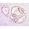
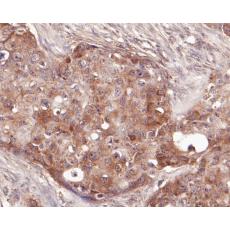
Fig7: Immunohistochemical analysis of paraffin-embedded human breast cancer tissue using anti-delta 1 Catenin/CAS antibody. The section was pre-treated using heat mediated antigen retrieval with Tris-EDTA buffer (pH 8.0-8.4) for 20 minutes.The tissues were blocked in 5% BSA for 30 minutes at room temperature, washed with ddH2O and PBS, and then probed with the primary antibody for 30 minutes at room temperature. The detection was performed using an HRP conjugated compact polymer system. DAB was used as the chromogen. Tissues were counterstained with hematoxylin and mounted with DPX.
Fig8: Immunohistochemical analysis of paraffin-embedded human kidney tissue using anti-delta 1 Catenin/CAS antibody. The section was pre-treated using heat mediated antigen retrieval with Tris-EDTA buffer (pH 8.0-8.4) for 20 minutes.The tissues were blocked in 5% BSA for 30 minutes at room temperature, washed with ddH2O and PBS, and then probed with the primary antibody for 30 minutes at room temperature. The detection was performed using an HRP conjugated compact polymer system. DAB was used as the chromogen. Tissues were counterstained with hematoxylin and mounted with DPX.
Fig9: Immunohistochemical analysis of paraffin-embedded mouse testis tissue using anti-delta 1 Catenin/CAS antibody. The section was pre-treated using heat mediated antigen retrieval with Tris-EDTA buffer (pH 8.0-8.4) for 20 minutes.The tissues were blocked in 5% BSA for 30 minutes at room temperature, washed with ddH2O and PBS, and then probed with the primary antibody for 30 minutes at room temperature. The detection was performed using an HRP conjugated compact polymer system. DAB was used as the chromogen. Tissues were counterstained with hematoxylin and mounted with DPX.
Fig10: Flow cytometric analysis of delta 1 Catenin/CAS was done on Siha cells. The cells were fixed, permeabilized and stained with the primary antibody (red). After incubation of the primary antibody at room temperature for an hour, the cells were stained with a Alexa Fluor 488-conjugated goat anti-rabbit IgG Secondary antibody at 1/500 dilution for 30 minutes.Unlabelled sample was used as a control (cells without incubation with primary antibody; black).
特别提示:本公司的所有产品仅可用于科研实验,严禁用于临床医疗及其他非科研用途!