Anti-ICAM1 antibody
-
概述
- 产品描述ICAM-1 is a member of the immunoglobulin superfamily, the superfamily of proteins including antibodies and T-cell receptors. The structure of ICAM-1 is characterized by heavy glycosylation, and the protein’s extracellular domain is composed of multiple loops created by disulfide bridges within the protein. ICAM-1 can be induced by interleukin-1 (IL-1) and tumor necrosis factor (TNF) and is expressed by the vascular endothelium, macrophages, and lymphocytes. ICAM-1 is a ligand for LFA-1 (integrin), a receptor found on leukocytes. More recently, ICAM-1 has been characterized as a site for the cellular entry of human rhinovirus. ICAM-1 and soluble ICAM-1 have antagonistic effects on the tight junctions forming the blood-testis barrier, thus playing a major role in spermatogenesis. ICAM-1 has been implicated in subarachnoid hemorrhage (SAH).
- 产品名称Anti-ICAM1 antibody
- 分子量90 kDa
- 种属反应性Human
- 验证应用WB,ICC,IHC-P,FC
- 抗体类型兔多抗
- 免疫原peptide
- 偶联Non-conjugated
-
性能
- 形态Liquid
- 浓度1 mg/mL.
- 存放说明Store at +4℃ after thawing. Aliquot store at -20℃ or -80℃. Avoid repeated freeze / thaw cycles.
- 存储缓冲液1*PBS (pH7.4), 0.2% BSA, 40% Glycerol. Preservative: 0.05% Sodium Azide.
- 亚型IgG
- 纯化方式Peptide affinity purified
- 亚细胞定位Cell membrane, Nucleus, Mitochondrion
- 其它名称Antigen identified by monoclonal antibody
BB2 antibody
BB 2 antibody
BB2 antibody
CD 54 antibody
CD_antigen=CD54 antibody
CD54 antibody
Cell surface glycoprotein P3.58 antibody
Human rhinovirus receptor antibody
ICAM 1 antibody
ICAM-1 antibody
ICAM1 antibody
ICAM1_HUMAN antibody
intercellular adhesion molecule 1 (CD54), human rhinovirus receptor antibody
Intercellular adhesion molecule 1 antibody
Major group rhinovirus receptor antibody
MALA 2 antibody
MALA2 antibody
MyD 10 antibody
MyD10 antibody
P3.58 antibody
Surface antigen of activated B cells, BB2 antibody
more
-
应用
WB: 1:500-1:1,000
ICC: 1:200
IHC-P: 1:200
FC: 1:50-1:100
-
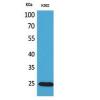
Fig1: Western blot analysis of ICAM1 on different cell lysates using anti- ICAM1 antibody at 1/500 dilution.
Positive control:
Lane 1: Raji
Lane 2: HUVEC
Lane 3: K562
Fig2: ICC staining of ICAM1 in HUVEC cells (green). Cells were fixed in paraformaldehyde, permeabilised with 0.25% Triton X100/PBS.
Fig3: Immunohistochemical analysis of paraffin-embedded human tonsil tissue using anti-ICAM1 antibody. Counter stained with hematoxylin.
Fig4: Flow cytometric analysis of Hela cells with ICAM1 antibody at 1/100 dilution (blue) compared with an unlabelled control (cells without incubation with primary antibody; red). Goat anti rabbit IgG (FITC) was used as the secondary antibody.
特别提示:本公司的所有产品仅可用于科研实验,严禁用于临床医疗及其他非科研用途!