Anti-Factor XIIIa antibody
-
概述
- 产品描述This gene encodes the coagulation factor XIII A subunit. Coagulation factor XIII is the last zymogen to become activated in the blood coagulation cascade. Plasma factor XIII is a heterotetramer composed of 2 A subunits and 2 B subunits. The A subunits have catalytic function, and the B subunits do not have enzymatic activity and may serve as plasma carrier molecules. Platelet factor XIII is composed of just 2 A subunits, which are identical to those of plasma origin. Upon cleavage of the activation peptide by thrombin and in the presence of calcium ion, the plasma factor XIII dissociates its B subunits and yields the same active enzyme, factor XIIIa, as platelet factor XIII. This enzyme acts as a transglutaminase to catalyze the formation of gamma-glutamyl-epsilon-lysine crosslinking between fibrin molecules, thus stabilizing the fibrin clot. It also crosslinks alpha-2-plasmin inhibitor, or fibronectin, to the alpha chains of fibrin. Factor XIII deficiency is classified into two categories: type I deficiency, characterized by the lack of both the A and B subunits; and type II deficiency, characterized by the lack of the A subunit alone. These defects can result in a lifelong bleeding tendency, defective wound healing, and habitual abortion.
- 产品名称Anti-Factor XIIIa antibody
- 分子量Predicted band size 83 kDa.
- 种属反应性Human
- 验证应用WB,IHC-P,FC
- 抗体类型兔多抗
- 免疫原Recombinant protein within human Factor XIIIa aa 200-500.
- 偶联Non-conjugated
-
性能
- 形态Liquid
- 浓度1 mg/ml.
- 存放说明Store at +4℃ after thawing. Aliquot store at -20℃. Avoid repeated freeze / thaw cycles.
- 存储缓冲液1*PBS (pH7.4), 0.2% BSA, 50% Glycerol. Preservative: 0.05% Sodium Azide.
- 亚型IgG
- 纯化方式Protein affinity purified.
- 亚细胞定位Cytoplasm, Secreted.
- 其它名称
- bA525O21.1 (coagulation factor XIII, A1 polypeptide) antibody
- Coagulation factor XIII A chain antibody
- Coagulation factor XIII A1 polypeptide antibody
more
-
应用
WB: 1:500-1:1000
IHC-P: 1:50-1:200
FC: 1:50-1:100
-
Fig1: Western blot analysis of Factor XIIIa on different lysates. Proteins were transferred to a PVDF membrane and blocked with 5% BSA in PBS for 1 hour at room temperature. The primary antibody was used in 5% BSA at room temperature for 2 hours. Goat Anti-Rabbit IgG - HRP Secondary Antibody (HA1001) at 1:5,000 dilution was used for 1 hour at room temperature.
Positive control:
Lane 1: human placenta tissue lysate
Lane 2: human liver tissue lysate
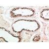
Fig2: Immunohistochemical analysis of paraffin-embedded human placenta tissue using anti-Factor XIIIa antibody. The section was pre-treated using heat mediated antigen retrieval with Tris-EDTA buffer (pH 8.0-8.4) for 20 minutes.The tissues were blocked in 5% BSA for 30 minutes at room temperature, washed with ddH2O and PBS, and then probed with the primary antibody for 30 minutes at room temperature. The detection was performed using an HRP conjugated compact polymer system. DAB was used as the chromogen. Tissues were counterstained with hematoxylin and mounted with DPX.
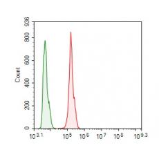
Fig3: Flow cytometric analysis of Factor XIIIa was done on Hela cells. The cells were fixed, permeabilized and stained with the primary antibody (red). After incubation of the primary antibody at room temperature for an hour, the cells were stained with a Alexa Fluor 488-conjugated goat anti-rabbit IgG Secondary antibody at 1/500 dilution for 30 minutes.Unlabelled sample was used as a control (cells without incubation with primary antibody; green).
特别提示:本公司的所有产品仅可用于科研实验,严禁用于临床医疗及其他非科研用途!