Anti-AFP antibody
-
概述
- 产品描述AFP is a protein normally produced by the fetal liver and is present in the fluid surrounding the fetus (amniotic fluid), and crosses the placenta into the mother's blood. AFP gene expression is regulated by the interactions between steroid hormone receptors and transcriptional factors in separate signal transduction pathways. AFP functions as a binding and transporting ligand and cell growth regulator. Elevated expression level of AFP has been implicated in colorectal, ovarian, pancreatic, testicular, and certain liver cancers. High levels of AFP are also seen in some diseases such as hepatitis and colitis.
- 产品名称买二赠二Anti-AFP antibody
- 分子量70 kDa
- 种属反应性Human
- 验证应用WB,ICC,IHC-P,FC
- 抗体类型兔多抗
- 免疫原peptide
- 偶联Non-conjugated
-
性能
- 形态Liquid
- 浓度1 mg/mL.
- 存放说明Store at +4℃ after thawing. Aliquot store at -20℃ or -80℃. Avoid repeated freeze / thaw cycles.
- 存储缓冲液1*PBS (pH7.4), 0.2% BSA, 40% Glycerol. Preservative: 0.05% Sodium Azide.
- 亚型IgG
- 纯化方式Peptide affinity purified
- 亚细胞定位Secreted
- 其它名称
- Afp antibody
- AFPD antibody
- Alpha fetoglobulin antibody
more
-
应用
WB: 1:1,000-1:2,000
ICC: 1:200
IHC-P: 1:200
FC: 1:100-1:200
-
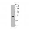
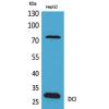
Fig1: Western blot analysis of AFP on different cell lysates using anti-AFP antibody at 1/1000 dilution.
Positive control:
Lane 1: HepG2
Lane 2: human lung
Lane 3: human liver
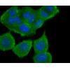
Fig2: ICC staining AFP in NCCIT cells (green). Cells were fixed in paraformaldehyde, permeabilised with 0.25% Triton X100/PBS.
Fig3: ICC staining AFP in HepG2 cells (green). Cells were fixed in paraformaldehyde, permeabilised with 0.25% Triton X100/PBS.
Fig4: Immunohistochemical analysis of paraffin-embedded human liver cancer tissue using anti-AFP antibody. Counter stained with hematoxylin.
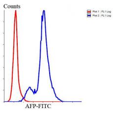
Fig5: Flow cytometric analysis of Jurkat cells with AFP antibody at 1/100 dilution (blue) compared with an unlabelled control (cells without incubation with primary antibody; red). Goat anti rabbit IgG (FITC) was used as the secondary antibody.
特别提示:本公司的所有产品仅可用于科研实验,严禁用于临床医疗及其他非科研用途!