


-

专业包装 正品保证
-

快乐服务 售后无忧
-

会员特权 优惠不断
-

个人信息 严格保护
| 货号 | 规格 | 可用库存 | 销售价(RMB) | 您的折扣价(RMB) | 购买数量 |
|---|
| 熔点: | |
|---|---|
| 密度: | |
| 储存条件: | -20℃ |
Anti-cMet antibody
产品描述c-Met (MET or MNNG HOS Transforming gene) is a proto-oncogene that encodes a protein known as hepatocyte growth factor receptor (HGFR). MET is a membrane receptor that is essential for embryonic development and wound healing. Hepatocyte growth factor (HGF) is the only known ligand of the MET receptor. MET is normally expressed by cells of epithelial origin, while expression of HGF is restricted to cells of mesenchymal origin. Upon HGF stimulation, MET induces several biological responses that collectively give rise to a program known as invasive growth. Abnormal MET activation in cancer correlates with poor prognosis, where aberrantly active MET triggers tumor growth, formation of new blood vessels (angiogenesis) that supply the tumor with nutrients, and cancer spread to other organs (metastasis). MET is deregulated in many types of human malignancies, including cancers of kidney, liver, stomach, breast, and brain.
产品名称Anti-cMet antibody
分子量153 kDa
种属反应性Human, Mouse, Rat
验证应用WB, ICC, IHC-P, FC
抗体类型兔多抗
免疫原Synthetic peptide within mouse cMet aa 650-690.
偶联Non-conjugated
形态Liquid
浓度1 mg/mL.
存放说明Store at +4℃ after thawing. Aliquot store at -20℃ or -80℃. Avoid repeated freeze / thaw cycles.
存储缓冲液1*PBS (pH7.4), 0.2% BSA, 40% Glycerol. Preservative: 0.05% Sodium Azide.
亚型IgG
纯化方式Peptide affinity purified
亚细胞定位Cell membrane.
其它名称
WB: 1:1,000
ICC: 1:200
IHC-P: 1:200
FC: 1:100-1:200

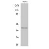
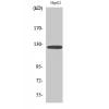
Fig1: Western blot analysis of cMet on different cell lysates using anti-cMet antibody at 1/1000 dilution.
Positive control:
Lane 1: Mouse liver
Lane 2: Mouse kidney
Lane 3: D3
Lane 4: MEF

Fig2: ICC staining cMet in N2A cells (green). Cells were fixed in paraformaldehyde, permeabilised with 0.25% Triton X100/PBS.

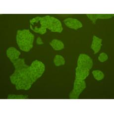
Fig3: ICC staining cMet in Hela cells (green). Cells were fixed in paraformaldehyde, permeabilised with 0.25% Triton X100/PBS.

Fig4: ICC staining cMet in HepG2 cells (green). Cells were fixed in paraformaldehyde, permeabilised with 0.25% Triton X100/PBS.

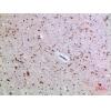
Fig5: Immunohistochemical analysis of paraffin-embedded mouse liver tissue using anti-cMet antibody. Counter stained with hematoxylin.

Fig6: Immunohistochemical analysis of paraffin-embedded mouse spleen tissue using anti-cMet antibody. Counter stained with hematoxylin.

Fig7: Immunohistochemical analysis of paraffin-embedded human colon cancer tissue using anti-cMet antibody. Counter stained with hematoxylin.

Fig8: Flow cytometric analysis of Hela cells with cMet antibody at 1/100 dilution (blue) compared with an unlabelled control (cells without incubation with primary antibody; red). Goat anti rabbit IgG (FITC) was used as the secondary antibody.
特别提示:本公司的所有产品仅可用于科研实验,严禁用于临床医疗及其他非科研用途!


