


-

专业包装 正品保证
-

快乐服务 售后无忧
-

会员特权 优惠不断
-

个人信息 严格保护
| 货号 | 规格 | 可用库存 | 销售价(RMB) | 您的折扣价(RMB) | 购买数量 |
|---|
| 熔点: | |
|---|---|
| 密度: | |
| 储存条件: | -20℃ |
Anti-CD31 antibody
产品描述PECAM-1 is found on the surface of platelets, monocytes, neutrophils, and some types of T-cells, and makes up a large portion of endothelial cell intercellular junctions. The encoded protein is a member of the immunoglobulin superfamily and is likely involved in leukocyte transmigration, angiogenesis, and integrin activation. CD31 is also expressed in certain tumors, including epithelioid hemangioendothelioma, epithelioid sarcoma-like hemangioendothelioma, other vascular tumors, histiocytic malignancies, and plasmacytomas. It is rarely found in some sarcomas, such as Kaposi's sarcoma and carcinomas. In immunohistochemistry, CD31 is used primarily to demonstrate the presence of endothelial cells in histological tissue sections. This can help to evaluate the degree of tumour angiogenesis, which can imply a rapidly growing tumor
产品名称Anti-CD31 antibody
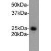
分子量82 kDa (Observed: 130 kDa)
种属反应性Human,Mouse
验证应用WB,ICC,IHC-P,FC
抗体类型兔多抗
免疫原This antibody is produced by immunizing rabbits with a synthetic peptide (KLH-coupled) corresponding to C-terminal residues of human CD31.
偶联Non-conjugated
形态Liquid
浓度1 mg/mL.
存放说明Store at +4℃ after thawing. Aliquot store at -20℃. Avoid repeated freeze / thaw cycles.
存储缓冲液1*PBS (pH7.4), 0.2% BSA, 40% Glycerol. Preservative: 0.05% Sodium Azide.
亚型IgG
纯化方式Peptide affinity purified
亚细胞定位Cell membrane
其它名称
WB: 1:1,000
ICC: 1:200
IHC-P: 1:200
FC: 1:100-1:200

Fig1: Western blot analysis of CD31 on different cell lysates using anti-CD31 antibody at 1/1000 dilution.

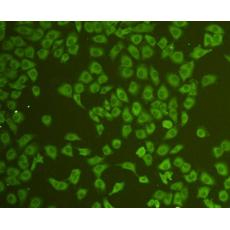
Fig2: ICC staining CD31 in Hela cells (green). Cells were fixed in paraformaldehyde, permeabilised with 0.25% Triton X100/PBS.

Fig3: ICC staining CD31 in HUVEC cells (green). Cells were fixed in paraformaldehyde, permeabilised with 0.25% Triton X100/PBS.

Fig4: ICC staining CD31 in NIH/3T3 cells (green). Cells were fixed in paraformaldehyde, permeabilised with 0.25% Triton X100/PBS.

Fig5: ICC staining CD31 in SW480 cells (green). Cells were fixed in paraformaldehyde, permeabilised with 0.25% Triton X100/PBS.

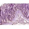
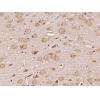
Fig6: Immunohistochemical analysis of paraffin-embedded human kidney tissue using anti-CD31 antibody. Counter stained with hematoxylin.

Fig7: Flow cytometric analysis of Jurkat cells with CD31 antibody at 1/100 dilution (blue) compared with an unlabelled control (cells without incubation with primary antibody; red). Goat anti rabbit IgG (FITC) was used as the secondary antibody.
特别提示:本公司的所有产品仅可用于科研实验,严禁用于临床医疗及其他非科研用途!


