Anti-Bcl2 antibody
-
概述
- 产品描述Suppresses apoptosis in a variety of cell systems including factor-dependent lymphohematopoietic and neural cells. Regulates cell death by controlling the mitochondrial membrane permeability. Appears to function in a feedback loop system with caspases. Inhibits caspase activity either by preventing the release of cytochrome c from the mitochondria and/or by binding to the apoptosis-activating factor (APAF-1). May attenuate inflammation by impairing NLRP1-inflammasome activation, hence CASP1 activation and IL1B release.
- 产品名称Anti-Bcl2 antibody
- 分子量22/26 kDa
- 种属反应性Human,Mouse
- 验证应用WB,ICC,FC
- 抗体类型兔多抗
- 免疫原Synthetic peptide within human BCL-2 20-100 aa.
- 偶联Non-conjugated
-
性能
- 形态Liquid
- 浓度1 mg/mL.
- 存放说明Store at +4℃ after thawing. Aliquot store at -20℃. Avoid repeated freeze / thaw cycles.
- 存储缓冲液1*PBS (pH7.4), 0.2% BSA, 50% Glycerol. Preservative: 0.05% Sodium Azide.
- 亚型IgG
- 纯化方式Peptide affinity purified.
- 亚细胞定位Mitochondrion. Endoplasmic reticulum. Nucleus.
- 其它名称Apoptosis regulator Bcl 2 antibody
Apoptosis regulator Bcl-2 antibody
Apoptosis regulator Bcl2 antibody
AW986256 antibody
B cell CLL/lymphoma 2 antibody
B cell leukemia/lymphoma 2 antibody
Bcl-2 antibody
Bcl2 antibody
BCL2_HUMAN antibody
C430015F12Rik antibody
D630044D05Rik antibody
D830018M01Rik antibody
Leukemia/lymphoma, B-cell, 2 antibody
Oncogene B-cell leukemia 2 antibody
PPP1R50 antibody
Protein phosphatase 1, regulatory subunit 50 antibody
more
-
应用
WB: 1:200-1:500
ICC: 1:50-1:200
FC: 1:50-1:100
-
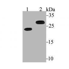
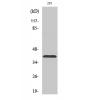
Fig1: Western blot analysis of Bcl2 on different lysates using anti-Bcl2 antibody at 1/200 dilution.
Positive control:
Lane 1: HL-60
Lane 2: Mouse colon
Fig2: ICC staining Bcl2 in LOVO cells (green). The nuclear counter stain is DAPI (blue). Cells were fixed in paraformaldehyde, permeabilised with 0.25% Triton X100/PBS.
Fig3: ICC staining Bcl2 in MCF-7 cells (green). The nuclear counter stain is DAPI (blue). Cells were fixed in paraformaldehyde, permeabilised with 0.25% Triton X100/PBS.
Fig4: ICC staining Bcl2 in SH-SY-5Y cells (green). The nuclear counter stain is DAPI (blue). Cells were fixed in paraformaldehyde, permeabilised with 0.25% Triton X100/PBS.
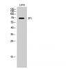
Fig5: Flow cytometric analysis of MCF-7 cells with Bcl2 antibody at 1/100 dilution (purple) compared with an unlabelled control (cells without incubation with primary antibody; yellow). Alexa Fluor 488-conjugated goat anti-rabbit IgG was used as the secondary antibody.
特别提示:本公司的所有产品仅可用于科研实验,严禁用于临床医疗及其他非科研用途!