


-

专业包装 正品保证
-

快乐服务 售后无忧
-

会员特权 优惠不断
-

个人信息 严格保护
| 别名: | CD86 | ||
|---|---|---|---|
| 适用物种: | Human,Mouse,Rat,Dog,Pig,Cow,Sheep | ||
| 验证应用: | WB,IHC-P,ICC/IF,FC | ||
| 种属: | 兔多抗 | ||
| 储存条件: | -20℃ | ||
|
| 货号 | 规格 | 可用库存 | 销售价(RMB) | 您的折扣价(RMB) | 购买数量 | ||||||
|---|---|---|---|---|---|---|---|---|---|---|---|
| ZY6906-01R-50 μl | 兔多抗 | 现货 | 1500 | ||||||||
| ZY6906-01R-100 μl | 兔多抗 | 现货 | 2500 | ||||||||
| 熔点: | |
|---|---|
| 密度: | |
| 储存条件: | -20℃ |
Anti-CD86 antibody
产品描述Receptor involved in the costimulatory signal essential for T-lymphocyte proliferation and interleukin-2 production, by binding CD28 or CTLA-4. May play a critical role in the early events of T-cell activation and costimulation of naive T-cells, such as deciding between immunity and anergy that is made by T-cells within 24 hours after activation. Isoform 2 interferes with the formation of CD86 clusters, and thus acts as a negative regulator of T-cell activation.
产品名称Anti-CD86 antibody
分子量31 KDa
种属反应性Human,Mouse,Rat,Dog,Pig,Cow,Sheep
验证应用WB,IHC-P,ICC/IF,FC
抗体类型兔多抗
免疫原KLH conjugated synthetic peptide derived from the middle of human CD86 140-175/313
偶联Non-conjugated
形态Liquid
浓度1 mg/mL.
存放说明Store at -20℃ for one year. Avoid repeated freeze/thaw cycles. The lyophilized antibody is stable at room temperature for at least one month and for greater than a year when kept at -20℃. When reconstituted in sterile pH 7.4 0.01M PBS or diluent of antibody the antibody is stable for at least two weeks at 2-4℃.
存储缓冲液0.01M TBS(pH7.4) with 1% BSA, 0.03% Proclin300 and 50% Glycerol.
亚型IgG
纯化方式affinity purified by Protein A
亚细胞定位Cell membrane; Single-pass type I membrane protein.
其它名称
WB:1:500-2000
IHC-P:1:400-800
FC:1μg/Test
IF:1:100-500
ICC/IF:1:100-500

Fig1: Tissue/cell: BV-2 cell; 4% Paraformaldehyde-fixed; Triton X-100 at room temperature for 20 min; Blocking buffer (normal goat serum, C-0005) at 37℃ for 20 min; Antibody incubation with (CD86) Polyclonal Antibody, Unconjugated 1:200, 90 minutes at 37℃; followed by a conjugated Goat Anti-Rabbit IgG antibody (bs-0295G-FITC) at 37℃ for 90 minutes, DAPI (5ug/ml, blue, C-0033) was used to stain the cell nuclei.

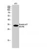
Fig2: Sample:
Lymph node(Mouse)Cell Lysate at 40 ug
Primary: Anti-CD86at 1/300 dilution
Secondary: IRDye800CW Goat Anti-Rabbit IgG at 1/20000 dilution
Predicted band size: 31 kD
Observed band size: 31 kD

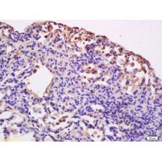
Fig3: Tissue/cell: rat lung tissue; 4% Paraformaldehyde-fixed and paraffin-embedded;
Antigen retrieval: citrate buffer ( 0.01M, pH 6.0 ), Boiling bathing for 15min; Block endogenous peroxidase by 3% Hydrogen peroxide for 30min; Blocking buffer (normal goat serum,C-0005) at 37℃ for 20 min;
Incubation: Anti-CD86/B7-2 Polyclonal Antibody, Unconjugated1:200, overnight at 4℃, followed by conjugation to the secondary antibody(SP-0023) and DAB(C-0010) staining

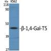
Fig4: Sample:
Brain(Rat) lysates at 30ug;
Heart(Rat) lysates, 30ug;
Primary: Anti-CD86/B7-2 at 1:200;
Secondary: HRP conjugated Goat Anti-Rabbit IgG(bs-0295G-HRP) at 1: 3000;
ECL excitated the fluorescence;
Predicted band size : 34kD
Observed band size : 34kD

Fig5: Blank control: U937(blue).
Primary Antibody: Rabbit Anti-CD86 antibody ), Dilution: 1μg in 100 μL 1X PBS containing 0.5% BSA;
Isotype Control Antibody: Rabbit IgG (orange) ,used under the same conditions.
Secondary Antibody: Goat anti-rabbit IgG-PE(white blue), Dilution: 1:200 in 1 X PBS containing 0.5% BSA.
Protocol
The cells were fixed with 2% paraformaldehyde (10 min).Primary antibody (, 1μg /1x10^6 cells) were incubated for 30 min on the ice, followed by 1 X PBS containing 0.5% BSA + 10% goat serum (15 min) to block non-specific protein-protein interactions. Then the Goat Anti-rabbit IgG/PE antibody was added into the blocking buffer mentioned above to react with the primary antibody at 1/200 dilution for 30 min on ice. Acquisition of 20,000 events was performed.
特别提示:本公司的所有产品仅可用于科研实验,严禁用于临床医疗及其他非科研用途!


