Anti-VPS4a antibody
-
概述
- 产品描述Involved in late steps of the endosomal multivesicular bodies (MVB) pathway. Recognizes membrane-associated ESCRT-III assemblies and catalyzes their disassembly, possibly in combination with membrane fission. Redistributes the ESCRT-III components to the cytoplasm for further rounds of MVB sorting. MVBs contain intraluminal vesicles (ILVs) that are generated by invagination and scission from the limiting membrane of the endosome and mostly are delivered to lysosomes enabling degradation of membrane proteins, such as stimulated growth factor receptors, lysosomal enzymes and lipids. In conjunction with the ESCRT machinery also appears to function in topologically equivalent membrane fission events, such as the terminal stages of cytokinesis and enveloped virus budding (HIV-1 and other lentiviruses). Involved in cytokinesis.
- 产品名称Anti-VPS4a antibody
- 分子量48 KDa
- 种属反应性Human,Mouse,Rat,Dog,Pig,Rabbit
- 验证应用WB,IHC-P
- 抗体类型兔多抗
- 免疫原KLH conjugated synthetic peptide derived from human VPS4a 351-437/437
- 偶联Non-conjugated
-
性能
- 形态Liquid
- 浓度1 mg/mL.
- 存放说明Store at -20℃ for one year. Avoid repeated freeze/thaw cycles. The lyophilized antibody is stable at room temperature for at least one month and for greater than a year when kept at -20℃. When reconstituted in sterile pH 7.4 0.01M PBS or diluent of antibody the antibody is stable for at least two weeks at 2-4℃.
- 存储缓冲液0.01M TBS(pH7.4) with 1% BSA, 0.03% Proclin300 and 50% Glycerol.
- 亚型IgG
- 纯化方式affinity purified by Protein A
- 亚细胞定位Prevacuolar compartment membrane; Peripheral membrane protein. Late endosome membrane; Peripheral membrane protein (Probable). Note=Membrane-associated in the prevacuolar endosomal compartment. Localizes to the midbody of dividing cells. Localized in two
- 其它名称hVPS4
SKD1
SKD1 homolog
SKD2
Vacuolar protein sorting 4 homolog A
vacuolar sorting protein 4
VPS4.
more
-
应用
WB:1:500-2000
IHC-P:1:400-800
-
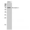
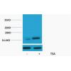
Fig1: Sample:
U-87MG(Human) Cell Lysate at 30 ug
NIH/3T3(Mouse) Cell Lysate at 30 ug
Primary: Anti-VPS4aat 1/500 dilution
Secondary: IRDye800CW Goat Anti-Rabbit IgG at 1/20000 dilution
Predicted band size: 48 kD
Observed band size: 48 kD
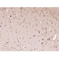
Fig2: Paraformaldehyde-fixed, paraffin embedded (Mouse brain); Antigen retrieval by boiling in sodium citrate buffer (pH6.0) for 15min; Block endogenous peroxidase by 3% hydrogen peroxide for 20 minutes; Blocking buffer (normal goat serum) at 37℃ for 30min; Antibody incubation with (VPS4a) Polyclonal Antibody, Unconjugated at 1:400 overnight at 4℃, followed by operating according to SP Kit(Rabbit) (sp-0023) instructionsand DAB staining.
特别提示:本公司的所有产品仅可用于科研实验,严禁用于临床医疗及其他非科研用途!