Anti-Progesterone Receptor antibody
-
概述
- 产品描述The effects of progesterone are mediated by two functionally different isoforms of the progesterone receptor, PR-A and PR-B, which are transcribed from distinct, estrogen-inducible promoters within a single copy of the PR gene. The first 164 amino acids of PR-B are absent in PR-A. Progesterone-bound PR-A and PR-B have different transcription activation properties. Specifically, PR-B functions as a transcriptional activator in most cell and promoter contexts, while PR-A is transcriptionally inactive and functions as a strong ligand-dependent transdominant repressor of steroid hormone receptor transcriptional activity. An inhibitory domain (ID), which maps to the amino terminus of the receptor, exists within both PR isoforms. Interestingly, the ID is functionally active only in PR-A and is necessary for steroid hormone transrepression by PR-A, suggesting that PR-A and PR-B may have different conformations in the cell.
- 产品名称Anti-Progesterone Receptor antibody
- 分子量99 kDa
- 种属反应性Human,Mouse
- 验证应用ICC,IHC-P
- 抗体类型兔多抗
- 免疫原Peptide
- 偶联Non-conjugated
-
性能
- 形态Liquid
- 浓度1 mg/mL.
- 存放说明Store at +4℃ after thawing. Aliquot store at -20℃ or -80℃. Avoid repeated freeze / thaw cycles.
- 存储缓冲液1*PBS (pH7.4), 0.2% BSA, 50% Glycerol. Preservative: 0.05% Sodium Azide.
- 亚型IgG
- 纯化方式Peptide affinity purified
- 亚细胞定位Cytoplasm. Nucleus.
- 其它名称NR3C3 antibody
Nuclear receptor subfamily 3 group C member 3 antibody
PGR antibody
PR antibody
PRA antibody
PRB antibody
PRGR_HUMAN antibody
Progesterone receptor antibody
Progestin receptor form A antibody
Progestin receptor form B antibody
more
-
应用
ICC: 1:500-1:1,000
IHC-P: 1:100-1:500
-
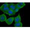
Fig1: ICC staining Progesterone Receptor in A549 cells (green). The nuclear counter stain is DAPI (blue). Cells were fixed in paraformaldehyde, permeabilised with 0.25% Triton X100/PBS.
Fig2: ICC staining Progesterone Receptor in Hela cells (green). The nuclear counter stain is DAPI (blue). Cells were fixed in paraformaldehyde, permeabilised with 0.25% Triton X100/PBS.
Fig3: ICC staining Progesterone Receptor in MCF-7 cells (green). The nuclear counter stain is DAPI (blue). Cells were fixed in paraformaldehyde, permeabilised with 0.25% Triton X100/PBS.
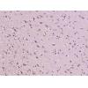
Fig4: Immunohistochemical analysis of paraffin-embedded human uterus tissue using anti-Progesterone Receptor antibody. Counter stained with hematoxylin.
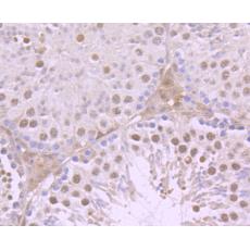
Fig5: Immunohistochemical analysis of paraffin-embedded mouse testis tissue using anti-Progesterone Receptor antibody. Counter stained with hematoxylin.
特别提示:本公司的所有产品仅可用于科研实验,严禁用于临床医疗及其他非科研用途!


