Anti-Mitofusin 2 antibody
-
概述
- 产品描述Mitofusin 1 (Mfn1) and mitofusin 2 (Mfn2) are homologs for the Drosophila protein fuzzy onion (Fzo). They are mitochondrial membrane proteins and are mediators of mitochondrial fusion. A GTPase domain is required for Mfn protein function but the molecular mechanisms of the GTPase-dependent reaction as well as the functional division of the two Mfn proteins are unknown. They are essential for embryonic development and may play a role in the pathobiology of obesity. Although the Mfn1 and Mfn2 genes are broadly expressed, they show different levels of expression in different tissues. Two Mfn1 transcripts are elevated in heart, while Mfn2 mRNA is abundantly expressed in heart and muscle tissue but present only at low levels in many other tissues. Mfn1 localizes to mitochondria and participates in at least two different high molecular weight protein complexes in a GTP-dependent manner. Purified recombinant Mfn1 exhibited approximately eightfold higher GTPase activity than Mfn2.
- 产品名称Anti-Mitofusin 2 antibody
- 分子量86 kDa
- 种属反应性Human,Rat
- 验证应用WB,ICC,FC
- 抗体类型兔多抗
- 免疫原Recombinant protein
- 偶联Non-conjugated
-
性能
- 形态Liquid
- 浓度1 mg/mL.
- 存放说明Store at +4℃ after thawing. Aliquot store at -20℃ or -80℃. Avoid repeated freeze / thaw cycles.
- 存储缓冲液1*PBS (pH7.4), 0.2% BSA, 50% Glycerol. Preservative: 0.05% Sodium Azide.
- 亚型IgG
- 纯化方式Protein affinity purified
- 亚细胞定位Mitochondrion.
- 其它名称CMT2A antibody
CMT2A2 antibody
CPRP 1 antibody
CPRP1 antibody
EC 3.6.5.- antibody
Fzo antibody
HSG antibody
hyperplasia suppressor gene antibody
Hypertension related protein 1 antibody
KIAA0214 antibody
MARF antibody
MFN 2 antibody
Mfn2 antibody
MFN2_HUMAN antibody
Mitochondrial assembly regulatory factor antibody
Mitofusin-2 antibody
Mitofusin2 antibody
Transmembrane GTPase MFN2 antibody
more
-
应用
WB: 1:500-1:1,000
ICC: 1:50-1:200
FC: 1:50-1:100
-
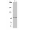
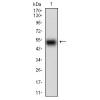
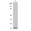
Fig1: Western blot analysis of Mitofusin 2 on Rat heart tissue lysates using anti-Mitofusin 2 antibody at 1/1,000 dilution.
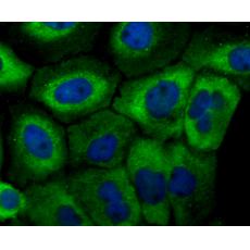
Fig2: ICC staining Mitofusin 2 in A431 cells (green). The nuclear counter stain is DAPI (blue). Cells were fixed in paraformaldehyde, permeabilised with 0.25% Triton X100/PBS.
Fig3: ICC staining Mitofusin 2 in LOVO cells (green). The nuclear counter stain is DAPI (blue). Cells were fixed in paraformaldehyde, permeabilised with 0.25% Triton X100/PBS.
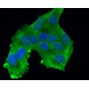
Fig4: ICC staining Mitofusin 2 in PC-3M cells (green). The nuclear counter stain is DAPI (blue). Cells were fixed in paraformaldehyde, permeabilised with 0.25% Triton X100/PBS.
Fig5: Flow cytometric analysis of Jurkat cells with Mitofusin 2 antibody at 1/100 dilution (red) compared with an unlabelled control (cells without incubation with primary antibody; black). Alexa Fluor 488-conjugated goat anti-rabbit IgG was used as the secondary antibody.
特别提示:本公司的所有产品仅可用于科研实验,严禁用于临床医疗及其他非科研用途!