Anti-Cullin-3 antibody
-
概述
- 产品描述Cullin proteins comprise a distinct family of mediators that participate in the selective targeting of proteins for ubiquitin (Ub)-mediated proteolysis. CUL-1, which is the mammalian homolog of yeast Cdc53, is an integral component of the E3 ubiquitin ligase complex designated SCF. The SCF (Skp1/CUL-1/F-box protein complex) consists of Skp1 associating with both CUL-1 and an F-box protein, such as Skp2, which determines the substrate specificity of the complex. CUL-1-mediated ubiquitination results in the degradation of cell cycle proteins cyclin D, p21 and cyclin E. Another cullin, CUL-3, facilitates the degradation of cyclin E independent of SCF activity, while CUL-2 associates with the tumor suppressing protein VHL and elongin B to form VBC complexes, which structurally resemble the SCF ligase. Proteolysis also occurs by way of CUL-4 associating with Nedd-8, a ubiquitin-like protein, where it too functions as an active component of a multifunctional E3 complex. CUL-5, also designated vasopressin-activated, calcium-mobilizing protein (VACM-1), is also included in the cullin family as it shares substantial sequence homology with CUL-1.
- 产品名称Anti-Cullin-3 antibody
- 分子量89 kDa
- 种属反应性Human,Mouse
- 验证应用WB,IHC-P,FC
- 抗体类型兔多抗
- 免疫原Peptide.
- 偶联Non-conjugated
-
性能
- 形态Liquid
- 浓度1 mg/mL.
- 存放说明Store at +4℃ after thawing. Aliquot store at -20℃ or -80℃. Avoid repeated freeze / thaw cycles.
- 存储缓冲液1*PBS (pH7.4), 0.2% BSA, 50% Glycerol. Preservative: 0.05% Sodium Azide.
- 亚型IgG
- 纯化方式Peptide affinity purified
- 亚细胞定位Golgi apparatus. Nucleus.
- 其它名称CUL 3 antibody
Cul-3 antibody
cul3 antibody
CUL3_HUMAN antibody
Cullin-3 antibody
Cullin3 antibody
KIAA0617 antibody
PHA2E antibody
more
-
应用
WB: 1:500-1:1,000
IHC-P: 1:50-1:200
FC: 1:50-1:100
-
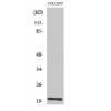
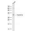
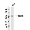
Fig1: Western blot analysis of Cullin 3 on different lysates using anti-Cullin 3 antibody at 1/1,000 dilution.
Positive control:
Lane 1: SH-SY5Y
Lane 2: 293T
Lane 3: Mouse testis tissue
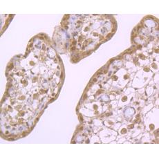
Fig2: Immunohistochemical analysis of paraffin-embedded human kidney tissue using anti-Cullin-3 antibody. Counter stained with hematoxylin.
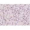
Fig3: Immunohistochemical analysis of paraffin-embedded human placenta tissue using anti-Cullin-3 antibody. Counter stained with hematoxylin.
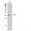
Fig4: Flow cytometric analysis of SH-SY5Y cells with Cullin 3 antibody at 1/100 dilution (red) compared with an unlabelled control (cells without incubation with primary antibody; black). Alexa Fluor 488-conjugated goat anti-rabbit IgG was used as the secondary antibody.
特别提示:本公司的所有产品仅可用于科研实验,严禁用于临床医疗及其他非科研用途!