


-

专业包装 正品保证
-

快乐服务 售后无忧
-

会员特权 优惠不断
-

个人信息 严格保护
| 货号 | 规格 | 可用库存 | 销售价(RMB) | 您的折扣价(RMB) | 购买数量 | ||||||
|---|---|---|---|---|---|---|---|---|---|---|---|
| ZY6901-30R-50 μl | 兔多抗 | 现货 | 1500 | ||||||||
| ZY6901-30R-100 μl | 兔多抗 | 现货 | 2500 | ||||||||
| 熔点: | |
|---|---|
| 密度: | |
| 储存条件: | -20℃ |
Anti-DOG1 antibody
产品描述ANO1 (anoctamin 1), also known as DOG1, ORAOV2, TAOS2 or TMEM16A, is a 986 amino acid multi-pass membrane protein that localizes to both the cell membrane and the cytoplasm and belongs to the anoctamin family. Expressed in a variety of tissues with highest expression in liver, gastrointestinal muscle and skeletal muscle, ANO1 functions as a calcium-activated chloride channel that is required for normal tracheal development. Human ANO1 shares 90% sequence identity with its mouse counterpart, suggesting a conserved role between species. ANO1 is present in breast, pancreatic, gastric, and uterine cancers, as well as in neck, ovarian and parathyroid tumors, suggesting a role for ANO1 in carcinogenesis. Three isoforms of ANO1 exist due to alternative splicing events.
产品名称Anti-DOG1 antibody
分子量114/97 kDa (Predicted band size)
种属反应性Human
验证应用WB,ICC,IHC-P,FC
抗体类型兔多抗
免疫原Synthetic peptide within C-terminal human CD43.
偶联Non-conjugated
形态Liquid
浓度1mg/mL
存放说明Store at +4℃ after thawing. Aliquot store at -20℃. Avoid repeated freeze / thaw cycles.
存储缓冲液1*PBS (pH7.4), 0.2% BSA, 50% Glycerol. Preservative: 0.05% Sodium Azide.
亚型IgG
纯化方式Peptide affinity purified.
亚细胞定位Cell membrane, cytoplasm.
其它名称
WB:1:500-1:1000
ICC:1:50-1:200
IHC-P:1:50-1:200
FC:1:50-1:100

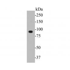
Fig1: Western blot analysis of DOG1 on PC-3M cell lysates. Proteins were transferred to a PVDF membrane and blocked with 5% BSA in PBS for 1 hour at room temperature. The primary antibody was used in 5% BSA at room temperature for 2 hours. Goat Anti-Rabbit IgG - HRP Secondary Antibody (HA1001) at 1:5,000 dilution was used for 1 hour at room temperature.

Fig2: ICC staining of DOG1 in A549 cells (green). Formalin fixed cells were permeabilized with 0.1% Triton X-100 in TBS for 10 minutes at room temperature and blocked with 1% Blocker BSA for 15 minutes at room temperature. Cells were probed with the primary antibody for 1 hour at room temperature, washed with PBS. Alexa Fluor®488 Goat anti-Rabbit IgG was used as the secondary antibody at 1/1,000 dilution. The nuclear counter stain is DAPI (blue).

Fig3: ICC staining of DOG1 in HepG2 cells (green). Formalin fixed cells were permeabilized with 0.1% Triton X-100 in TBS for 10 minutes at room temperature and blocked with 1% Blocker BSA for 15 minutes at room temperature. Cells were probed with the primary antibody for 1 hour at room temperature, washed with PBS. Alexa Fluor®488 Goat anti-Rabbit IgG was used as the secondary antibody at 1/1,000 dilution. The nuclear counter stain is DAPI (blue).

Fig4: Immunohistochemical analysis of paraffin-embedded human liver carcinoma tissue using anti-DOG1 antibody. The section was pre-treated using heat mediated antigen retrieval with Tris-EDTA buffer (pH 8.0-8.4) for 20 minutes.The tissues were blocked in 5% BSA for 30 minutes at room temperature, washed with ddH2O and PBS, and then probed with the primary antibody for 30 minutes at room temperature. The detection was performed using an HRP conjugated compact polymer system. DAB was used as the chromogen. Tissues were counterstained with hematoxylin and mounted with DPX.

Fig5: Immunohistochemical analysis of paraffin-embedded human seminal pouch tissue using anti-DOG1 antibody. The section was pre-treated using heat mediated antigen retrieval with Tris-EDTA buffer (pH 8.0-8.4) for 20 minutes.The tissues were blocked in 5% BSA for 30 minutes at room temperature, washed with ddH2O and PBS, and then probed with the primary antibodyfor 30 minutes at room temperature. The detection was performed using an HRP conjugated compact polymer system. DAB was used as the chromogen. Tissues were counterstained with hematoxylin and mounted with DPX.

Fig6: Flow cytometric analysis of DOG1 was done on A549 cells. The cells were fixed, permeabilized and stained with the primary antibody (red). After incubation of the primary antibody at room temperature for an hour, the cells were stained with a Alexa Fluor 488-conjugated goat anti-rabbit IgG Secondary antibody at 1/1000 dilution for 30 minutes.Unlabelled sample was used as a control (cells without incubation with primary antibody; black).
特别提示:本公司的所有产品仅可用于科研实验,严禁用于临床医疗及其他非科研用途!


