
![Anti-NME1 antibody [C6-B2]](images/202012/goods_img/92786_G_1608033562589.jpg)


-

专业包装 正品保证
-

快乐服务 售后无忧
-

会员特权 优惠不断
-

个人信息 严格保护
| 货号 | 规格 | 可用库存 | 销售价(RMB) | 您的折扣价(RMB) | 购买数量 | ||||||
|---|---|---|---|---|---|---|---|---|---|---|---|
| ZY6709-02M-50 μl | 小鼠单抗 | 现货 | 1500 | ||||||||
| ZY6709-02M-100 μl | 小鼠单抗 | 现货 | 2500 | ||||||||
| 熔点: | |
|---|---|
| 密度: | |
| 储存条件: | -20℃ |
Anti-NME1 antibody [C6-B2]
产品描述The nm23 protein is implicated in a variety of biological processes, including suppressing metastasis, phytochrome responses in plants and regulating differentiation. Nm23-H1, nm23-H2 and nm23-H2, members of the nm23 family, are putative metastasis suppressor genes that encode nucleoside diphosphate kinase (NDPK) A and B. NDPKs form oligomers, which are distributed in both the soluble and particulate fractions of cells. Upon cAMP-induced differentiation of rat cells, nm23 proteins show a differential interaction with intermediate filaments. Both isoforms also associate with GFAP in differentiated cells. nm23 proteins and their mutants are localized predominantly in the mitochondria. Nm23 mutants are unable to inhibit differentiation and promote apoptosis as a results of defective protein-protein interactions in the mitochondria. Nm23-H1, nm23-H2 and nm23-H3 are indicators of a poor prognosis in human hematopoietic malignancies, and as a high expression of nm23-H1 and -H2 is positively correlated with histological
产品名称Anti-NME1 antibody [C6-B2]
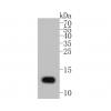
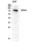
分子量17 kDa
种属反应性Human
验证应用WB,ICC,IHC-P,FC
抗体类型小鼠单抗
免疫原Recombinant protein
偶联Non-conjugated
形态Liquid
浓度2 mg/mL.
存放说明Store at +4℃ after thawing. Aliquot store at -20℃ or -80℃. Avoid repeated freeze / thaw cycles.
存储缓冲液1*TBS (pH7.4), 1%BSA, Preservative: 0.05% Sodium Azide.
亚型IgG1
纯化方式Protein A purified.
亚细胞定位Cytoplasm. Nucleus.
其它名称
AWD antibody
AWD, drosophila, homolog of antibody
GAAD antibody
WB: 1:500-1:1,000
ICC: 1:50-1:200
IHC-P: 1:50-1:200
FC: 1:50-1:200

Fig1: Western blot analysis of NME1 on NME1-hIgGFc transfected HEK293 (2) cell lysate using anti-NME1 antibody at 1/1,000 dilution.

Fig2: ICC staining NME1 (green) and Actin filaments (red) in Hela cells. The nuclear counter stain is DAPI (blue). Cells were fixed in paraformaldehyde, permeabilised with 0.25% Triton X100/PBS.

Fig3: Immunohistochemical analysis of paraffin-embedded human placenta tissue using anti-NME1 antibody. Counter stained with hematoxylin.

Fig4: Flow cytometric analysis of Jurkat cells with NME1 antibody at 1/100 dilution (green) compared with an unlabelled control (cells without incubation with primary antibody; purple).
特别提示:本公司的所有产品仅可用于科研实验,严禁用于临床医疗及其他非科研用途!


