Anti-TIE1 antibody [D2-D12]
-
概述
- 产品描述Receptor tyrosine kinases play key roles in signal transduction across cell surfaces in biological systems, including the vascular system. These receptors comprise a large and diverse family of catalytically related proteins that, on the basis of sequence and structural similarities, can be divided into several different evolutionary subfamilies. The cloning and characterization of Tie-1 (also designated Tie), a novel human endothelial cell surface receptor tyrosine kinase, has been reported. The extracellular domain of the predicted Tie-1 protein product has an unusual multidomain structure consisting of a cluster of three epidermal growth factor homology motifs localized between two immunoglobulin-like loops, which are followed by three fibronectin type III repeats next to the transmembrane region. An additional member of this family has been identified as Tie-2 (also designated Tek). Tie-1 and Tie-2 have been shown to be encoded by distinct genes and to represent members of a new class of receptor tyrosine kinases.
- 产品名称Anti-TIE1 antibody [D2-D12]
- 分子量125 kDa
- 种属反应性Human
- 验证应用WB,IHC-P
- 抗体类型小鼠单抗
- 免疫原Recombinant protein
- 偶联Non-conjugated
-
性能
- 形态Liquid
- 浓度2 mg/mL.
- 存放说明Store at +4℃ after thawing. Aliquot store at -20℃ or -80℃. Avoid repeated freeze / thaw cycles.
- 存储缓冲液1*TBS (pH7.4), 1%BSA, 40%Glycerol. Preservative: 0.05% Sodium Azide.
- 亚型IgG1
- 纯化方式Protein A purified.
- 亚细胞定位Cell membrane.
- 其它名称
- JKT 14 antibody
- JTK14 antibody
- TIE antibody
more
-
应用
WB: 1:500-1:2,000
IHC-P: 1:200-1:1,000
-
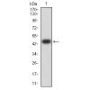
Fig1: Western blot analysis of TIE1 on human TIE1 recombinant protein using anti-TIE1 antibody at 1/1,000 dilution.
Fig2: Western blot analysis of TIE1 on HEK293 (1) and RAN-hIgGFc transfected HEK293 (2) cell lysate using anti-TIE1 antibody at 1/1,000 dilution.
Fig3: Western blot analysis of TIE1 on HepG2 cell lysate using anti-TIE1 antibody at 1/1,000 dilution.
Fig4: Immunohistochemical analysis of paraffin-embedded human ovarian cancer tissue using anti-CD68 antibody. Counter stained with hematoxylin.
Fig5: Immunohistochemical analysis of paraffin-embedded human kidney tissue using anti-CD68 antibody. Counter stained with hematoxylin.
特别提示:本公司的所有产品仅可用于科研实验,严禁用于临床医疗及其他非科研用途!


![Anti-TIE1 antibody [D2-D12]](images/202012/goods_img/92623_G_1607499195896.jpg)


