Anti-CBX1 antibody [B8-D7]
-
概述
- 产品描述Chromatin assembly factor-1 (CAF-1) is a multisubunit protein complex that comprises three polypeptide subunits known as p150, p60, and p48. CAF-1 is a nucleosome assembly factor that deposits newly synthesized and acetylated histones H3/H4 into nascent chromatin during DNA replication. The p150 subunit of CAF-1 also supports the maintenance of heterochromatin, which requires the synthesis of both new histones and heterochromatin proteins and their orderly assembly during DNA replication. Heterochromatin is characterized as densely coiled chromatin that generally replicates late during S phase, has a low gene density, and contains large blocks of repetitive DNA that is relatively inaccessible to DNA-modifying reagents. In late S phase, p150 directly associates with heterochromatin associated proteins 1 (HP1α, HP1β and HP1γ). As cells prepare for mitosis, CAF-1 p150 and some HP1 progressively dissociate from heterochromatin, coinciding with the phosphorylation of Histone H3. The HP1 proteins reassociate with chromatin at the end of mitosis, as Histone H3 is dephosphorylated.
- 产品名称Anti-CBX1 antibody [B8-D7]
- 分子量21 kDa
- 种属反应性Human
- 验证应用WB,ICC,IHC-P,FC
- 抗体类型小鼠单抗
- 免疫原Recombinant protein
- 偶联Non-conjugated
-
性能
- 形态Liquid
- 浓度2 mg/mL.
- 存放说明Store at +4℃ after thawing. Aliquot store at -20℃ or -80℃. Avoid repeated freeze / thaw cycles.
- 存储缓冲液1*TBS (pH7.4), 1%BSA, 40%Glycerol. Preservative: 0.05% Sodium Azide.
- 亚型IgG1
- 纯化方式Protein A purified.
- 亚细胞定位Nucleus
- 其它名称
- CBX 1 antibody
- CBX antibody
- Cbx1 antibody
more
-
应用
WB: 1:500-1:2,000
ICC: 1:50-1:200
IHC-P: 1:50-1:200
FC: 1:50-1:100
-
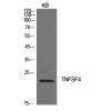
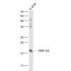
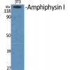
Fig1: Western blot analysis of CBX1 on human CBX1 recombinant protein using anti-CBX1 antibody at 1/1,000 dilution.
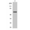
Fig2: Western blot analysis of CBX1 on HEK293 (1) and CBX1-hIgGFc transfected HEK293 (2) cell lysate using anti-CBX1 antibody at 1/1,000 dilution.
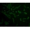
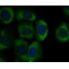
Fig3: ICC staining CBX1 (green) and Actin filaments (red) in Hela cells. The nuclear counter stain is DAPI (blue). Cells were fixed in paraformaldehyde, permeabilised with 0.25% Triton X100/PBS.
Fig4: Immunohistochemical analysis of paraffin-embedded human cervical cancer tissue using anti-CBX1 antibody. Counter stained with hematoxylin.
Fig5: Immunohistochemical analysis of paraffin-embedded human ovarian cancer tissue using anti-CBX1 antibody. Counter stained with hematoxylin.
Fig6: Immunohistochemical analysis of paraffin-embedded human colon cancer tissue using anti-CBX1 antibody. Counter stained with hematoxylin.
Fig7: Flow cytometric analysis of Hela cells with CBX1 antibody at 1/100 dilution (green) compared with an unlabelled control (cells without incubation with primary antibody; red).
特别提示:本公司的所有产品仅可用于科研实验,严禁用于临床医疗及其他非科研用途!

![Anti-CBX1 antibody [B8-D7]](images/202012/goods_img/92617_G_1607498823455.jpg)