Anti-EMD antibody [A8-F5]
-
概述
- 产品描述Emerin is believed to be a member of the nuclear lamina associated protein family. It is ubiquitously expressed and localized to the nuclear membrane in normal cells. Mutations of the gene that encodes emerin result in the X-linked recessive disease Emery-Dreyfuss muscular dystrophy (EDMD), which is characterized by slowly progressing contractures, skeletal muscle wasting and cardiomyopathy. Research has demonstrated that the lack of emerin expression is one cause of EDMD. Emerin is involved in the association of the nuclear membrane with the lamina, and is localized specifically to desmosomes and fasciae adherentes in the heart. This may account for conduction defects in patients with EDMD.
- 产品名称Anti-EMD antibody [A8-F5]
- 分子量29 kDa
- 种属反应性Human
- 验证应用WB,ICC,IHC-P,FC
- 抗体类型小鼠单抗
- 免疫原Recombinant protein
- 偶联Non-conjugated
-
性能
- 形态Liquid
- 浓度2 mg/mL.
- 存放说明Store at +4℃ after thawing. Aliquot store at -20℃ or -80℃. Avoid repeated freeze / thaw cycles.
- 存储缓冲液1*TBS (pH7.4), 1%BSA, 40%Glycerol. Preservative: 0.05% Sodium Azide.
- 亚型IgG1
- 纯化方式Protein A purified.
- 亚细胞定位Nucleus inner membrane. Nucleus outer membrane
- 其它名称
- EDMD antibody
- Emd antibody
- EMD_HUMAN antibody
more
-
应用
WB: 1:500-1:2,000
ICC: 1:50-1:200
IHC-P: 1:50-1:200
FC: 1:50-1:100
-
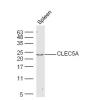
Fig1: Western blot analysis of Emerin on human Emerin recombinant protein using anti- Emerin antibody at 1/1,000 dilution.
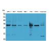
Fig2: Western blot analysis of Emerin on HEK293 (1) and Emerin -hIgGFc transfected HEK293 (2) cell lysate using anti- Emerin antibody at 1/1,000 dilution.
Fig3: ICC staining Emerin (green) and Actin filaments (red) in Hela cells. The nuclear counter stain is DAPI (blue). Cells were fixed in paraformaldehyde, permeabilised with 0.25% Triton X100/PBS.
Fig4: Immunohistochemical analysis of paraffin-embedded human ovarian cancer tissue using anti- Emerin antibody. Counter stained with hematoxylin.
Fig5: Flow cytometric analysis of A549 cells with Emerin antibody at 1/100 dilution (green) compared with an unlabelled control (cells without incubation with primary antibody; red).
Fig6: Flow cytometric analysis of K562 cells with Emerin antibody at 1/100 dilution (green) compared with an unlabelled control (cells without incubation with primary antibody; red).
特别提示:本公司的所有产品仅可用于科研实验,严禁用于临床医疗及其他非科研用途!

![Anti-EMD antibody [A8-F5]](images/202012/goods_img/92610_G_1607498469944.jpg)