Anti-RAD52 antibody [E11-E7]
-
概述
- 产品描述Rad52 family members (Rad50, Rad51B/C/D, Rad52, Rad54, MRE11) mediate DNA double-strand break repair (DSBR) for DNA damage that otherwise could cause cell death, mutation or neoplastic transformation. Rad51 (RECA, BRCC5) interacts with BRCA1 and BRCA2 to influence subcellular localization and cellular response to DNA damage. BRCA2 inactivation may be a key event leading to genomic instability and tumorigenesis from deregulation of Rad51. Rad52 forms a heptameric ring that binds single-stranded DNA ends and catalyzes DNA-DNA interaction necessary for the annealing of complementary strands. Rad52 can interact with Rad51. Rad54A of the DEAD-like helicase superfamily binds to double-strand DNA and induces a DNA topological change, which is thought to facilitate homologous DNA pairing, and stimulate DNA recombination. Rad54B of the DEAD-like helicase superfamily binds to double-stranded DNA and displays ATPase activity in the presence of DNA. Rad54B is abundant in testis and spleen, and mutations of this gene occur in primary lymphoma and colon cancer.
- 产品名称Anti-RAD52 antibody [E11-E7]
- 分子量46 kDa
- 种属反应性Human
- 验证应用WB,ICC,FC
- 抗体类型小鼠单抗
- 免疫原Recombinant protein
- 偶联Non-conjugated
-
性能
- 形态Liquid
- 浓度2 mg/mL.
- 存放说明Store at +4℃ after thawing. Aliquot store at -20℃ or -80℃. Avoid repeated freeze / thaw cycles.
- 存储缓冲液1*TBS (pH7.4), 1%BSA, 40%Glycerol. Preservative: 0.05% Sodium Azide.
- 亚型IgG1
- 纯化方式Protein A purified.
- 亚细胞定位Nucleus.
- 其它名称
- DNA repair protein RAD52 antibody
- DNA repair protein RAD52 antibody
- DNA repair protein RAD52 homolog antibody
more
-
应用
WB: 1:1,000-1:2,000
ICC: 1:50-1:200
FC: 1:50-1:100
-
Fig1: Western blot analysis of RAD52 on human RAD52 recombinant protein using anti-RAD52 antibody at 1/1,000 dilution.
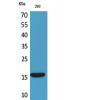
Fig2: Western blot analysis of RAD52 on HEK293 (1) and RAD52-hIgGFc transfected HEK293 (2) cell lysate using anti-RAD52 antibody at 1/1,000 dilution.
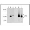
Fig3: Western blot analysis of RAD52 on different cell lysate using anti-RAD52 antibody at 1/1,000 dilution.
Positive control: Line1: HepG2 Line2: MCF-7 Line3: MCF-7 Line4: C6
Fig4: ICC staining RAD52 (green) and Actin filaments (red) in Hela cells. The nuclear counter stain is DAPI (blue). Cells were fixed in paraformaldehyde, permeabilised with 0.25% Triton X100/PBS.
Fig5: Flow cytometric analysis of MCF-7 cells with LAMP2 antibody at 1/100 dilution (green) compared with an unlabelled control (cells without incubation with primary antibody; red).
特别提示:本公司的所有产品仅可用于科研实验,严禁用于临床医疗及其他非科研用途!

![Anti-RAD52 antibody [E11-E7]](images/202012/goods_img/92555_G_1607493898263.jpg)