Anti-IL-31 antibody [8-C3]
-
概述
- 产品描述IL-31 is a T cell cytokine that is preferentially produced by T helper type 2 cells. IL-31 signals through a heterodimeric receptor composed of the IL-31 receptor (IL-31R) and the oncostatin M receptor (OSM). This receptor complex recruits JAK1, JAK2, Stat1, Stat3 and Stat5 signaling pathways, as well as the PI3 kinase/AKT cascade. SHP-2 and Shc adapter molecules are also recruited and contribute to an increased activation of the MAP kinase pathway in response to IL-31. Overexpression of IL-31 in mice results in pruritus and skin dermatitis resembling human atopic dermatitis (AD). Comparisons between skin from patients with AD and healthy skin showed IL-31R expression at higher levels on epidermal keratinocytes in AD samples. Infiltrating cells, more numerous in skin from patients with AD compared with that of healthy individuals, expressed IL-31 mRNA. IL-31 may participate in the cause of itch sensation and promote scratching behavior in NC/Nga mice with atopic dermatitis, and may represent a novel target for antipruritic drug development.
- 产品名称Anti-IL-31 antibody [8-C3]
- 分子量18 kDa
- 种属反应性Human
- 验证应用WB,IHC-P,ICC
- 抗体类型小鼠单抗
- 免疫原Recombinant full length protein corresponding to human IL18.
- 偶联Non-conjugated
-
性能
- 形态Liquid
- 浓度2 mg/mL.
- 存放说明Store at +4℃ after thawing. Aliquot store at -20℃. Avoid repeated freeze / thaw cycles.
- 存储缓冲液1*PBS (pH7.4), 0.2% BSA, 50% Glycerol. Preservative: 0.05% Sodium Azide.
- 亚型IgG3
- 纯化方式Protein affinity purified.
- 亚细胞定位Secreted.
- 其它名称
- IL 31 antibody
- IL-31 antibody
- IL31 antibody
more
-
应用
WB: 1:500
ICC: 1:100
IHC-P: 1:50-1:200
-
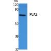
Fig1: Western blot analysis of IL-31 on recombinant protein lysate using anti-IL-31 antibody at 1/500 dilution.
Fig2: ICC staining IL-31 (green) in A431 cells. The nuclear counter stain is DAPI (blue). Cells were fixed in paraformaldehyde, permeabilised with 0.25% Triton X100/PBS.
Fig3: ICC staining IL-31 (green) in LOVO cells. The nuclear counter stain is DAPI (blue). Cells were fixed in paraformaldehyde, permeabilised with 0.25% Triton X100/PBS.
Fig4: ICC staining IL-31 (green) in MCF-7 cells. The nuclear counter stain is DAPI (blue). Cells were fixed in paraformaldehyde, permeabilised with 0.25% Triton X100/PBS.
Fig5: Immunohistochemical analysis of paraffin-embedded human tonsil tissue using anti-IL-31 antibody. Counter stained with hematoxylin.
Fig6: Immunohistochemical analysis of paraffin-embedded human spleen tissue using anti-IL-31 antibody. Counter stained with hematoxylin.
特别提示:本公司的所有产品仅可用于科研实验,严禁用于临床医疗及其他非科研用途!

![Anti-IL-31 antibody [8-C3]](images/202012/goods_img/92542_G_1607424564678.jpg)