Anti-YKL-40 / CHI3L1 antibody [10C1]
-
概述
- 产品描述Carbohydrate-binding lectin with a preference for chitin. Has no chitinase activity. May play a role in tissue remodeling and in the capacity of cells to respond to and cope with changes in their environment. Plays a role in T-helper cell type 2 (Th2) inflammatory response and IL-13-induced inflammation, regulating allergen sensitization, inflammatory cell apoptosis, dendritic cell accumulation and M2 macrophage differentiation. Facilitates invasion of pathogenic enteric bacteria into colonic mucosa and lymphoid organs. Mediates activation of AKT1 signaling pathway and subsequent IL8 production in colonic epithelial cells. Regulates antibacterial responses in lung by contributing to macrophage bacterial killing, controlling bacterial dissemination and augmenting host tolerance. Also regulates hyperoxia-induced injury, inflammation and epithelial apoptosis in lung.
- 产品名称Anti-YKL-40 / CHI3L1 antibody [10C1]
- 分子量43 kDa
- 种属反应性Human, Mouse
- 验证应用WB,IHC-P,ICC
- 抗体类型小鼠单抗
- 免疫原Synthetic peptide within Human YKL-40 / CHI3L1 aa 140-160.
- 偶联Non-conjugated
-
性能
- 形态Liquid
- 浓度2 mg/mL.
- 存放说明Store at +4℃ after thawing. Aliquot store at -20℃. Avoid repeated freeze / thaw cycles.
- 存储缓冲液1*PBS (pH7.4), 0.2% BSA, 50% Glycerol. Preservative: 0.05% Sodium Azide.
- 亚型IgG1
- 纯化方式Protein G purified.
- 亚细胞定位Cytoplasm, Endoplasmic reticulum, Secreted.
- 其它名称
- 39 kDa synovial protein antibody
- ASRT7 antibody
- Cartilage glycoprotein 39 antibody
more
-
应用
WB:1:500-1:1,000
IHC-P:1:50-1:200
FC:1:50-1:100
-
Fig1: Western blot analysis of YKL-40 / CHI3L1 on THP-1 cell lysates. Proteins were transferred to a PVDF membrane and blocked with 5% BSA in PBS for 1 hour at room temperature. The primary antibody was used in 5% BSA at room temperature for 2 hours. Goat Anti-Rabbit IgG - HRP Secondary Antibody (HA1001) at 1:5,000 dilution was used for 1 hour at room temperature.
Fig2: Immunohistochemical analysis of paraffin-embedded human tonsil tissue using anti-YKL-40 / CHI3L1 antibody. The section was pre-treated using heat mediated antigen retrieval with Tris-EDTA buffer (pH 8.0-8.4) for 20 minutes.The tissues were blocked in 5% BSA for 30 minutes at room temperature, washed with ddH2O and PBS, and then probed with the antibody at 1/50 dilution, for 30 minutes at room temperature and detected using an HRP conjugated compact polymer system. DAB was used as the chrogen. Counter stained with hematoxylin and mounted with DPX.
Fig3: Immunohistochemical analysis of paraffin-embedded human liver tissue using anti-YKL-40 / CHI3L1 antibody. The section was pre-treated using heat mediated antigen retrieval with sodium citrate buffer (pH 6.0) for 20 minutes. The tissues were blocked in 5% BSA for 30 minutes at room temperature, washed with ddH2O and PBS, and then probed with the antibody at 1/200 dilution, for 30 minutes at room temperature and detected using an HRP conjugated compact polymer system. DAB was used as the chrogen. Counter stained with hematoxylin and mounted with DPX.
Fig4: Immunohistochemical analysis of paraffin-embedded mouse lung tissue using anti-YKL-40 / CHI3L1 antibody. The section was pre-treated using heat mediated antigen retrieval with Tris-EDTA buffer (pH 8.0-8.4) for 20 minutes.The tissues were blocked in 5% BSA for 30 minutes at room temperature, washed with ddH2O and PBS, and then probed with the antibody at 1/200 dilution, for 30 minutes at room temperature and detected using an HRP conjugated compact polymer system. DAB was used as the chrogen. Counter stained with hematoxylin and mounted with DPX.
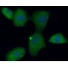
Fig5: Flow cytometric analysis of YKL-40 / CHI3L1 was done on HepG2 cells. The cells were fixed, permeabilized and stained with YKL-40 / CHI3L1 antibody at 1/100 dilution (red) compared with an unlabelled control (cells without incubation with primary antibody; black). After incubation of the primary antibody on room temperature for an hour, the cells was stained with a Alexa Fluor™ 488-conjugated goat anti-mouse IgG Secondary antibody at 1/500 dilution for 30 minutes.
特别提示:本公司的所有产品仅可用于科研实验,严禁用于临床医疗及其他非科研用途!


![Anti-YKL-40 / CHI3L1 antibody [10C1]](images/202012/goods_img/92457_G_1607330749714.jpg)


