Anti-CASK antibody [A1C7]
-
概述
- 产品描述Peripheral plasma membrane protein CASK is a protein that in humans is encoded by the CASK gene. This gene is also known by several other names: CMG 2 (CAMGUK protein 2), calcium/calmodulin-dependent serine protein kinase 3 and membrane-associated guanylate kinase 2.Multidomain scaffolding protein with a role in synaptic transmembrane protein anchoring and ion channel trafficking. Contributes to neural development and regulation of gene expression via interaction with the transcription factor TBR1. Binds to cell-surface proteins, including amyloid precursor protein, neurexins and syndecans. May mediate a link between the extracellular matrix and the actin cytoskeleton via its interaction with syndecan and with the actin/spectrin-binding protein 4.1.
- 产品名称Anti-CASK antibody [A1C7]
- 分子量105 kDa
- 种属反应性Human,Mouse,Rat
- 验证应用WB,IHC-P,FC
- 抗体类型小鼠单抗
- 免疫原Recombinant protein within Human CASK aa 320-550.
- 偶联Non-conjugated
-
性能
- 形态Liquid
- 浓度2 mg/ml.
- 存放说明Store at +4℃ after thawing. Aliquot store at -20℃. Avoid repeated freeze / thaw cycles.
- 存储缓冲液1*PBS (pH7.4), 0.2% BSA, 50% Glycerol. Preservative: 0.05% Sodium Azide.
- 亚型IgG1
- 纯化方式Protein A purified
- 亚细胞定位Cell membrane, Cytoplasm, Membrane, Nucleus.
- 其它名称
- CAGH39 antibody
- Caki antibody
- Calcium/calmodulin dependent serine protein kinase antibody
more
-
应用
WB:1:500-1:1000
IHC-P:1:50-1:200
FC:1:50-1:100
-
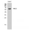
Fig1: Western blot analysis of CASK on different lysates. Proteins were transferred to a PVDF membrane and blocked with 5% BSA in PBS for 1 hour at room temperature. The primary antibody ( was used in 5% BSA at room temperature for 2 hours. Goat Anti-Mouse IgG - HRP Secondary Antibody (HA1006) at 1:5,000 dilution was used for 1 hour at room temperature.
Positive control:
Lane 1: Siha cell lysate
Lane 2: A549 cell lysate
Fig2: Immunohistochemical analysis of paraffin-embedded rat large intestine tissue using anti-CASK antibody. The section was pre-treated using heat mediated antigen retrieval with Tris-EDTA buffer (pH 8.0-8.4) for 20 minutes.The tissues were blocked in 5% BSA for 30 minutes at room temperature, washed with ddH2O and PBS, and then probed with the primary antibody for 30 minutes at room temperature. The detection was performed using an HRP conjugated compact polymer system. DAB was used as the chromogen. Tissues were counterstained with hematoxylin and mounted with DPX.
Fig3: Immunohistochemical analysis of paraffin-embedded human prostate cancer tissue using anti-CASK antibody. The section was pre-treated using heat mediated antigen retrieval with Tris-EDTA buffer (pH 8.0-8.4) for 20 minutes.The tissues were blocked in 5% BSA for 30 minutes at room temperature, washed with ddH2O and PBS, and then probed with the primary antibod for 30 minutes at room temperature. The detection was performed using an HRP conjugated compact polymer system. DAB was used as the chromogen. Tissues were counterstained with hematoxylin and mounted with DPX.
Fig4: Immunohistochemical analysis of paraffin-embedded mouse brain tissue using anti-CASK antibody. The section was pre-treated using heat mediated antigen retrieval with Tris-EDTA buffer (pH 8.0-8.4) for 20 minutes.The tissues were blocked in 5% BSA for 30 minutes at room temperature, washed with ddH2O and PBS, and then probed with the primary antibody for 30 minutes at room temperature. The detection was performed using an HRP conjugated compact polymer system. DAB was used as the chromogen. Tissues were counterstained with hematoxylin and mounted with DPX.
Fig5: Flow cytometric analysis of CASK was done on SW620 cells. The cells were fixed, permeabilized and stained with the primary antibody (red). After incubation of the primary antibody at room temperature for an hour, the cells were stained with a Alexa FluorTM488 Goat anti-Mouse IgG Secondary antibody at 1/500 dilution for 30 minutes.Unlabelled sample was used as a control (cells without incubation with primary antibody; black).
特别提示:本公司的所有产品仅可用于科研实验,严禁用于临床医疗及其他非科研用途!

![Anti-CASK antibody [A1C7]](images/202012/goods_img/92256_G_1607170646218.jpg)