Anti-SHC1 antibody [7C-7F]
-
概述
- 产品描述Growth factor triggering of protein tyrosine kinase receptors induces signals that cascade to the nucleus activating mitogenic, as well as other, responses. Critical components of this process include adapter proteins such as Shc and IRS-1 that lack detectable catalytic activity. These are immediate substrates of receptor tyrosine kinase activity and serve to physically link activated receptors to downstream signaling components. Whereas Shc has been implicated in signaling by diverse receptor families, IRS-1 serves primarily as the major insulin receptor substrate (4-7). Shc also participates in insulin signaling by linking the insulin receptor to Ras by forming complexes with the adapter protein GRB2 and Sos independently of IRS-1. A protein immunologically related to IRS-1, originally designated 4PS and now known as IRS-2, was shown to become highly tyrosine phosphorylated in response to IL-4 or IGF-1 in cells lacking IRS-1. An additional member of this family of signaling intermediates, Shb, is a SH2-containing protein with characteristic proline-rich domains.
- 产品名称Anti-SHC1 antibody [7C-7F]
- 分子量63 kDa
- 种属反应性Human,Mouse
- 验证应用WB,ICC,FC
- 抗体类型小鼠单抗
- 免疫原Recombinant protein
- 偶联Non-conjugated
-
性能
- 形态Liquid
- 浓度2 mg/mL.
- 存放说明Store at +4℃ after thawing. Aliquot store at -20℃ or -80℃. Avoid repeated freeze / thaw cycles.
- 存储缓冲液1*TBS (pH7.4), 1%BSA, 40%Glycerol. Preservative: 0.05% Sodium Azide.
- 亚型IgG1
- 纯化方式Protein A purified.
- 亚细胞定位Cytoplasm, Mitochondrion matrix.
- 其它名称
- FLJ26504 antibody
- p66 antibody
- p66SHC antibody
more
-
应用
WB: 1:500-1:2,000
ICC: 1:50-1:200
IHC-P: 1:50-1:200
FC: 1:50-1:100
-
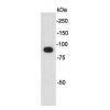
Fig1: Western blot analysis of SHC1 on human SHC1 recombinant protein using anti-SHC1 antibody at 1/1,000 dilution.
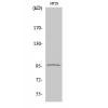
Fig2: Western blot analysis of SHC1 on HEK293 (1) and SHC1-hIgGFc transfected HEK293 (2) cell lysate using anti-SHC1 antibody at 1/1,000 dilution.
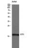
Fig3: Western blot analysis of SHC1 on NIH/3T3 cell lysate using anti-SHC1 antibody at 1/1,000 dilution.
Fig4: ICC staining SHC1 (green) and Actin filaments (red) in A431 cells. The nuclear counter stain is DAPI (blue). Cells were fixed in paraformaldehyde, permeabilised with 0.25% Triton X100/PBS.
Fig5: Flow cytometric analysis of NIH/3T3 cells with SHC1 antibody at 1/100 dilution (green) compared with an unlabelled control (cells without incubation with primary antibody; red).
特别提示:本公司的所有产品仅可用于科研实验,严禁用于临床医疗及其他非科研用途!

![Anti-SHC1 antibody [7C-7F]](images/202012/goods_img/92164_G_1606988220106.jpg)