Anti-Sequestosome-1 antibody
-
概述
- 产品描述Sequestosome 1 (SQSTM1, p62) is a ubiquitin binding protein involved in cell signaling, oxidative stress and autophagy. It was first identified as a protein that binds to the SH2 domain of p56Lck and independently found to interact with PKCζ. SQSTM1 was subsequently found to interact with ubiquitin, providing a scaffold for several signaling proteins and triggering degradation of proteins through the proteasome or lysosome. Interaction between SQSTM1 and TRAF6 leads to the K63-linked polyubiquitination of TRAF6 and subsequent activation of the NF-κB pathway. It may play a role in titin/TTN downstream signaling in muscle cells and regulate signaling cascades through ubiquitination. P62 may be involved in cell differentiation, apoptosis, immune response and regulation of K+ channels.
- 产品名称Anti-Sequestosome-1 antibody
- 分子量62 kDa
- 种属反应性Human,Mouse,Rat
- 验证应用WB,ICC,IHC-P,FC
- 抗体类型小鼠单抗
- 免疫原Synthetic peptide (KLH-coupled) within human SQSTM1 40-100 aa.
- 偶联Non-conjugated
-
性能
- 形态Liquid
- 浓度2 mg/mL.
- 存放说明Store at +4℃ after thawing. Aliquot store at -20℃. Avoid repeated freeze / thaw cycles.
- 存储缓冲液1*PBS (pH7.4), 0.2% BSA, 40% Glycerol. Preservative: 0.05% Sodium Azide.
- 亚型IgG1
- 纯化方式Peptide affinity purified.
- 亚细胞定位Cytoplasm, Nucleus
- 其它名称
- A170 antibody
- DMRV antibody
- EBI 3 associated protein of 60 kDa antibody
more
-
应用
WB: 1:500-1:1,000
ICC: 1:200
IHC-P: 1:200
FC: 1 μg per 1 x 10⁶ cells
-
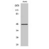
Fig1: Western blot analysis of SQSTM1 on different cell lysates using anti-SQSTM1 antibody at 1/500 dilution.
Positive control:
Lane 1: Hela
Lane 2: HepG2
Fig2: ICC staining SQSTM1 in HepG2 cells (red). Cells were fixed in paraformaldehyde, permeabilised with 0.25% Triton X100/PBS.
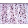
Fig3: Immunohistochemical analysis of paraffin-embedded mouse spleen tissue using anti- SQSTM1 antibody. Counter stained with hematoxylin.
Fig4: Immunohistochemical analysis of paraffin-embedded mouse prostate tissue using anti- SQSTM1 antibody. Counter stained with hematoxylin.
Fig5: Immunohistochemical analysis of paraffin-embedded mouse pancreas tissue using anti-SQSTM1 antibody. Counter stained with hematoxylin.
Fig6: Flow cytometric analysis of Hela cells with SQSTM1 antibody at 1/50 dilution (blue) compared with an unlabelled control (cells without incubation with primary antibody; red). Goat anti mouse IgG (FITC) was used as the secondary antibody.
特别提示:本公司的所有产品仅可用于科研实验,严禁用于临床医疗及其他非科研用途!