Anti-GAL4 antibody
-
概述
- 产品描述Gal4 is a modular protein consisting broadly of a DNA-binding domain and an activation domain. The UAS to which GAL4 binds is CGG-N11-CCG, where N can be any base.[6] Although GAL4 is a yeast protein not normally present in other organisms it has been shown to work as a transcription activator in a variety of organisms such as Drosophila, and human cells, highlighting that the same mechanisms for gene expression have been conserved over the course of evolution. GAL4 is then only expressed in cells where the driver gene is usually active. In turn, GAL4 should only activate gene transcription where a UAS has been introduced. For example, by fusing a gene encoding a visible marker like GFP (Green Fluorescent Protein) the expression pattern of the driver genes can be determined. GAL4 and the UAS are very useful for studying gene expression in Drosophila as they are not normally present and their expression does not interfere with other processes in the cell. For example, GAL4/UAS-regulated transgenes in Drosophila have been used to alter glial expression to produce arrhythmic behavior in a known rhythmic circadian output called pigment dispersing factor (PDF). However, some research has indicated that over-expression of GAL4 in Drosophila can have side-effects, probably relating to immune and stress responses to what is essentially an alien protein.
- 产品名称Anti-GAL4 antibody
- 分子量36 kDa
- 种属反应性Human,Mouse,Rat
- 验证应用WB,IHC-P,FC
- 抗体类型兔多抗
- 免疫原Recombinant protein corresponding to N-terminal Human GAL4.
- 偶联Non-conjugated
-
性能
- 形态Liquid
- 浓度1 mg/ml.
- 存放说明Store at +4℃ after thawing. Aliquot store at -20℃. Avoid repeated freeze / thaw cycles.
- 存储缓冲液1*PBS (pH7.4), 0.2% BSA, 50% Glycerol. Preservative: 0.05% Sodium Azide.
- 亚型IgG
- 纯化方式Protein affinity purified.
- 亚细胞定位Cytosol.
- 其它名称
- Antigen NY CO 27 antibody
- Antigen NY-CO-27 antibody
- Antigen NYCO27 antibody
more
-
应用
WB: 1:500-1:1,000
IHC-P: 1:50-1:200
FC: 1:50-1:100
-
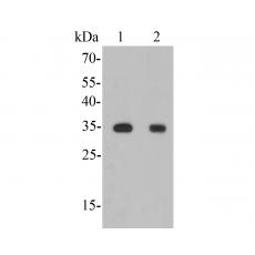
Fig1: Western blot analysis of GAL4 on different lysates. Proteins were transferred to a PVDF membrane and blocked with 5% BSA in PBS for 1 hour at room temperature. The primary antibody was used in 5% BSA at room temperature for 2 hours. Goat Anti-Rabbit IgG - HRP Secondary Antibody (HA1001) at 1:5,000 dilution was used for 1 hour at room temperature.
Positive control:
Lane 1: Human small intestine tissue lysate
Lane 2: Mouse colon tissue lysate
Fig2: Immunohistochemical analysis of paraffin-embedded rat large intestine tissue using anti-GAL4 antibody. The section was pre-treated using heat mediated antigen retrieval with Tris-EDTA buffer (pH 8.0-8.4) for 20 minutes.The tissues were blocked in 5% BSA for 30 minutes at room temperature, washed with ddH2O and PBS, and then probed with the primary antibody for 30 minutes at room temperature. The detection was performed using an HRP conjugated compact polymer system. DAB was used as the chromogen. Tissues were counterstained with hematoxylin and mounted with DPX.
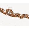
Fig3: Immunohistochemical analysis of paraffin-embedded human appendix tissue using anti-GAL4 antibody. The section was pre-treated using heat mediated antigen retrieval with Tris-EDTA buffer (pH 8.0-8.4) for 20 minutes.The tissues were blocked in 5% BSA for 30 minutes at room temperature, washed with ddH2O and PBS, and then probed with the primary antibody for 30 minutes at room temperature. The detection was performed using an HRP conjugated compact polymer system. DAB was used as the chromogen. Tissues were counterstained with hematoxylin and mounted with DPX.
Fig4: Immunohistochemical analysis of paraffin-embedded mouse colon tissue using anti-GAL4 antibody. The section was pre-treated using heat mediated antigen retrieval with Tris-EDTA buffer (pH 8.0-8.4) for 20 minutes.The tissues were blocked in 5% BSA for 30 minutes at room temperature, washed with ddH2O and PBS, and then probed with the primary antibody for 30 minutes at room temperature. The detection was performed using an HRP conjugated compact polymer system. DAB was used as the chromogen. Tissues were counterstained with hematoxylin and mounted with DPX.
Fig5: Flow cytometric analysis of GAL4 was done on SHSY5Y cells. The cells were fixed, permeabilized and stained with the primary antibody (purple). After incubation of the primary antibody at room temperature for an hour, the cells were stained with a Alexa Fluor 488-conjugated goat anti-rabbit IgG Secondary antibody at 1/500 dilution for 30 minutes. Unlabelled sample was used as a control (cells without incubation with primary antibody; yellow).
特别提示:本公司的所有产品仅可用于科研实验,严禁用于临床医疗及其他非科研用途!