Anti-FBXL2 antibody
-
概述
- 产品描述Calcium-activated substrate recognition component of a SCF (SKP1-cullin-F-box protein) E3 ubiquitin-protein ligase complex which mediates the ubiquitination and subsequent proteasomal degradation of target proteins. Unlike many F-box proteins, FBXL2 doesn't seem to target phosphodegron within its substrates but rather calmodulin-binding motifs. Targets PCYT1A for its monoubiquitination and degradation, this is antagonized by calmodulin (By similarity). Targets the cyclins CCND2 and CCND3 for polyubiquitination and degradation, leading to cell-cycle arrest in G(0), also antagonized by calmodulin. Binds to hepatitis C virus non-structural protein 5A (NS5A) in a reaction crucial for hepatitis C virus RNA replication.
- 产品名称Anti-FBXL2 antibody
- 分子量49 kDa
- 种属反应性Human,Mouse,Rat
- 验证应用WB,ICC,IHC-P
- 抗体类型兔多抗
- 免疫原Recombinant protein
- 偶联Non-conjugated
-
性能
- 形态Liquid
- 浓度1 mg/mL.
- 存放说明Store at +4℃after thawing. Aliquot store at -20℃or -80℃. Avoid repeated freeze / thaw cycles.
- 存储缓冲液1*PBS (pH7.4), 0.2% BSA, 50% Glycerol. Preservative: 0.05% Sodium Azide.
- 亚型IgG
- 纯化方式Protein A purified.
- 亚细胞定位Membrane.
- 其它名称
- DKFZP564P0622 antibody
- F box and leucine rich repeat protein 2 antibody
- F box protein containing leucine rich repeats antibody
more
-
应用
WB: 1:1,000-1:10,000
ICC: 1:50-1:200
IHC-P: 1:50-1:200
-
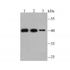
Fig1: Western blot analysis of FBXL2 on different lysates using anti-FBXL2 antibody at 1/1,000 dilution.
Positive control:
Lane 1: Mouse liver tissue Lane 2: Mouse heart tissue Lane 3: SiHa
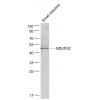
Fig2: Western blot analysis of FBXL2 on rat liver tissue lysate using anti-FBXL2 antibody at 1/10,000 dilution.
Fig3: ICC staining FBXL2 in A431 cells (green). The nuclear counter stain is DAPI (blue). Cells were fixed in paraformaldehyde, permeabilised with 0.25% Triton X100/PBS.
Fig4: ICC staining FBXL2 in HUVEC cells (green). The nuclear counter stain is DAPI (blue). Cells were fixed in paraformaldehyde, permeabilised with 0.25% Triton X100/PBS.
Fig5: ICC staining FBXL2 in SH-SY5Y cells (green). The nuclear counter stain is DAPI (blue). Cells were fixed in paraformaldehyde, permeabilised with 0.25% Triton X100/PBS.
Fig6: Immunohistochemical analysis of paraffin-embedded human liver tissue using anti-FBXL2 antibody. Counter stained with hematoxylin.
Fig7: Immunohistochemical analysis of paraffin-embedded human kidney tissue using anti-FBXL2 antibody. Counter stained with hematoxylin.
Fig8: Immunohistochemical analysis of paraffin-embedded human pancreas tissue using anti-FBXL2 antibody. Counter stained with hematoxylin.
Fig9: Immunohistochemical analysis of paraffin-embedded mouse brain tissue using anti-FBXL2 antibody. Counter stained with hematoxylin.
特别提示:本公司的所有产品仅可用于科研实验,严禁用于临床医疗及其他非科研用途!