Anti-eIF-4E antibody
-
概述
- 产品描述The initiation of protein synthesis in eukaryotic cells is regulated by interactions between protein initiation factors and RNA molecules. The eukaryotic initiation complex eIF4F exists in vitro as a trimeric complex of eIF4E, eIF4A and eIF4G. Together, the complex allows ribosome binding to mRNA by inducing the unwinding of mRNA secondary structures. eIF4E binds to the mRNA "cap" during an early step in the initiation of protein synthesis. eIF4A acts as an ATP-dependent RNA helicase. eIF4G acts as a bridge between eIF4E, eIF4A and the eIF3 complex.
- 产品名称Anti-eIF-4E antibody
- 分子量25 kDa
- 种属反应性Human,Mouse,Rat
- 验证应用WB,ICC,IHC-P,FC
- 抗体类型兔多抗
- 免疫原Peptide
- 偶联Non-conjugated
-
性能
- 形态Liquid
- 浓度1 mg/mL.
- 存放说明Store at +4℃ after thawing. Aliquot store at -20℃ or -80℃. Avoid repeated freeze / thaw cycles.
- 存储缓冲液1*PBS (pH7.4), 0.2% BSA, 50% Glycerol. Preservative: 0.05% Sodium Azide.
- 亚型IgG
- 纯化方式Peptide affinity purified
- 亚细胞定位Cytoplasm.
- 其它名称
- AUTS19 antibody
- CBP antibody
- eIF 4E antibody
more
-
应用
WB: 1:500-1:1,000
ICC: 1:50-1:200
IHC-P: 1:50-1:100
FC: 1:50-1:100
-
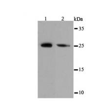
Fig1: Western blot analysis of eIF-4E on different cell lysates using anti-eIF-4E antibody at 1/500 dilution.
Positive control:
Lane 1: PC-12
Lane 2: NIH-3T3
Fig2: ICC staining eIF-4E in A431 cells (green). The nuclear counter stain is DAPI (blue). Cells were fixed in paraformaldehyde, permeabilised with 0.25% Triton X100/PBS.
Fig3: ICC staining eIF-4E in Hela cells (green). The nuclear counter stain is DAPI (blue). Cells were fixed in paraformaldehyde, permeabilised with 0.25% Triton X100/PBS.
Fig4: ICC staining eIF-4E in SK-Br-3 cells (green). The nuclear counter stain is DAPI (blue). Cells were fixed in paraformaldehyde, permeabilised with 0.25% Triton X100/PBS.
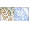
Fig5: Immunohistochemical analysis of paraffin-embedded human colon cancer tissue using anti-eIF-4E antibody. Counter stained with hematoxylin.
Fig6: Immunohistochemical analysis of paraffin-embedded mouse colon tissue using anti-eIF-4E antibody. Counter stained with hematoxylin.
Fig7: Immunohistochemical analysis of paraffin-embedded mouse brain tissue using anti-eIF-4E antibody. Counter stained with hematoxylin.
Fig8: Immunohistochemical analysis of paraffin-embedded mouse hippocampus tissue using anti-eIF-4E antibody. Counter stained with hematoxylin.
Fig9: Flow cytometric analysis of 293T cells with eIF-4E antibody at 1/100 dilution (red) compared with an unlabelled control (cells without incubation with primary antibody; black).
特别提示:本公司的所有产品仅可用于科研实验,严禁用于临床医疗及其他非科研用途!