Anti-PDGF Receptor beta antibody
-
概述
- 产品描述Platelet-derived growth factor (PDGF) is a mitogen for mesenchyme- and glia-derived cells. PDGF consists of two chains, A and B, which dimerize to form functionally distinct isoforms, PGDF-AA, PDGF-AB and PDGF-BB. These three isoforms bind with different affinities to two receptor types, PDGFR-α and -β, which are endowed with protein tyrosine kinase domains. PDGFR-α can bind to both A and B subunits of PDGF, while PDGFR- PDGF-AA induces the dimerization of αα and αβ and PDGF-BB induces the formation of three types of PDGF-AB induces dimerization of αα and αβ and PDGF-BB induces the formation of three types A dimlist, αα, αβ and ββ. Translocation of the PDGFR-β gene with the Tel gene is linked to chronic myelomonocytic leukemia (CMML), a myelodysplastic syndrome, and demonstrate the oncogenic potential of the PDGF receptors.
- 产品名称Anti-PDGF Receptor beta antibody
- 分子量175 kDa
- 种属反应性Human,Mouse,Rat
- 验证应用WB,ICC,IHC-P,FC
- 抗体类型兔多抗
- 免疫原Peptide
- 偶联Non-conjugated
-
性能
- 形态Liquid
- 浓度1 mg/mL.
- 存放说明Store at +4℃ after thawing. Aliquot store at -20℃ or -80℃. Avoid repeated freeze / thaw cycles.
- 存储缓冲液1*PBS (pH7.4), 0.2% BSA, 50% Glycerol. Preservative: 0.05% Sodium Azide.
- 亚型IgG
- 纯化方式Peptide affinity purified
- 亚细胞定位Membrane.
- 其它名称
- Beta platelet derived growth factor receptor antibody
- Beta-type platelet-derived growth factor receptor antibody
- CD 140B antibody
more
-
应用
ICC: 1:50-1:200
IHC-P: 1:50-1:200
FC: 1:50-1:100
WB: 1:500
-
Fig1: ICC staining PDGF Receptor beta in Hela cells (green). The nuclear counter stain is DAPI (blue). Cells were fixed in paraformaldehyde, permeabilised with 0.25% Triton X100/PBS.
Fig2: ICC staining PDGF Receptor beta in HepG2 cells (green). The nuclear counter stain is DAPI (blue). Cells were fixed in paraformaldehyde, permeabilised with 0.25% Triton X100/PBS.
Fig3: ICC staining PDGF Receptor beta in MCF-7 cells (green). The nuclear counter stain is DAPI (blue). Cells were fixed in paraformaldehyde, permeabilised with 0.25% Triton X100/PBS.
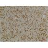
Fig4: Immunohistochemical analysis of paraffin-embedded human spleen tissue using anti-PDGF Receptor beta antibody. Counter stained with hematoxylin.
Fig5: Immunohistochemical analysis of paraffin-embedded mouse brain tissue using anti-PDGF Receptor beta antibody. Counter stained with hematoxylin.
Fig6: Flow cytometric analysis of NIH-3T3 cells with PDGF Receptor beta antibody at 1/100 dilution (red) compared with an unlabelled control (cells without incubation with primary antibody; black).
特别提示:本公司的所有产品仅可用于科研实验,严禁用于临床医疗及其他非科研用途!