Anti-GSK3 beta antibody
-
概述
- 产品描述Glycogen synthase kinase 3, or GSK-3, is a serine/threonine, proline-directed kinase involved in a diverse array of signaling pathways, including glycogen synthesis and cellular adhesion, and has been implicated in Alzheimer’s disease. Two forms of GSK-3, designated GSK-3α and GSK-3β, have been identified and differ in their subcellular localization. Tau, a microtubule-binding protein which serves to stabilize microtubules in growing axons, is found to be hyper-phosphorylated in paired helical filaments (PHF), the major fibrous component of neurofibrillary lesions associated with Alzheimer’s disease. Hyperphosphorylation of Tau is thought to be the critical event leading to the assembly of PHF. Six Tau protein isoforms have been identified, all of which are phosphorylated by GSK-3. This presents the possibility that miscues in GSK-3 signaling contribute to the onset of Alzheimer’s disease.
- 产品名称Anti-GSK3 beta antibody
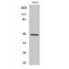
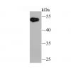
- 分子量46 kDa
- 种属反应性Human,Mouse,Rat
- 验证应用ICC,IHC-P,FC
- 抗体类型兔多抗
- 免疫原peptide
- 偶联Non-conjugated
-
性能
- 形态Liquid
- 浓度1 mg/mL.
- 存放说明Store at +4℃ after thawing. Aliquot store at -20℃ or -80℃. Avoid repeated freeze / thaw cycles.
- 存储缓冲液1*PBS (pH7.4), 0.2% BSA, 40% Glycerol. Preservative: 0.05% Sodium Azide.
- 亚型IgG
- 纯化方式Peptide affinity purified
- 亚细胞定位Cytoplasm, Nucleus, Cell membrane
- 其它名称
- Glycogen Synthase Kinase 3 Beta antibody
- Glycogen synthase kinase-3 beta antibody
- GSK 3 beta antibody
more
-
应用
ICC: 1:50-1:200
IHC-P: 1:50-1:200
FC: 1:50-1:100
-
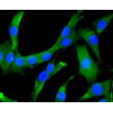
Fig1: Immunocytochemical staining of SHG-44 cells using anti-GSK3 beta rabbit polyclonal antibody.
Fig2: Immunocytochemical staining of MCF-7 cells using anti-GSK3 beta rabbit polyclonal antibody.
Fig3: Immunocytochemical staining of SW480 cells using anti-GSK3 beta rabbit polyclonal antibody.
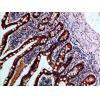
Fig4: Immunohistochemical analysis of paraffin- embedded human colon cancer tissue using anti-GSK3 beta rabbit polyclonal antibody.
Fig5: Immunohistochemical analysis of paraffin- embedded human breast carcinoma tissue using anti-GSK3 beta rabbit polyclonal antibody.
Fig6: Flow cytometric analysis of Jurkat cells with GSK3 beta antibody at 1/50 dilution (blue) compared with an unlabelled control (cells without incubation with primary antibody; red). Alexa Fluor 488-conjugated Goat anti mouse IgG was used as the secondary antibody.
特别提示:本公司的所有产品仅可用于科研实验,严禁用于临床医疗及其他非科研用途!


