Anti-ESD antibody
-
概述
- 产品描述ESD (esterase D) is also known as S-formylglutathione hydrolase and is a 282 amino acid protein that is a member of the esterase D family. ESD is highly expressed in placenta, kidney, liver and erythrocytes, and is localized to the cytoplasm, as well as to cytoplasmic vesicles. The main function of ESD is to detoxify formaldehyde while providing energy. Formaldehyde is oxidized by ADH5 which yields S-formylglutathione. ESD then catalyzes the hydrolysis of S-formylglutathione to the reduced forms of formic acid and glutathione. In addition, ESD hydrolyzes a variety of different neutral ester substrates and can act as a carboxylesterase. ESD may also act as a cysteine hydrolase which is inactivated by thiol alkylating agents. ESD gene polymorphism can lead to reduced enzymatic activity which may cause susceptibility to many conditions, including toxic liver cirrhosis, retinoblastoma, obesity and autism.
- 产品名称Anti-ESD antibody
- 分子量31 kDa
- 种属反应性Human,Mouse
- 验证应用WB,ICC,IHC-P
- 抗体类型兔多抗
- 免疫原Recombinant protein
- 偶联Non-conjugated
-
性能
- 形态Liquid
- 浓度1 mg/mL.
- 存放说明Store at +4℃after thawing. Aliquot store at -20℃or -80℃. Avoid repeated freeze / thaw cycles.
- 存储缓冲液1*PBS (pH7.4), 0.2% BSA, 40% Glycerol. Preservative: 0.05% Sodium Azide.
- 亚型IgG
- 纯化方式Peptide affinity purified
- 亚细胞定位Cytoplasm. Cytoplasmic vesicle.
- 其它名称
- EC 3.1.2.12 antibody
- Es-10 antibody
- Es10 antibody
more
-
应用
WB: 1:500-1:2000
ICC: 1:50-1:200
IHC-P: 1:50-1:200
-
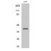
Fig1: Western blot analysis of ESD on K562 cell (1) and mouse intestine tissue (2) lysate using anti-ESD antibody at 1/1,000 dilution.
Fig2: ICC staining ESD in Hela cells (green). The nuclear counter stain is DAPI (blue). Cells were fixed in paraformaldehyde, permeabilised with 0.25% Triton X100/PBS.
Fig3: ICC staining ESD in LOVO cells (green). The nuclear counter stain is DAPI (blue). Cells were fixed in paraformaldehyde, permeabilised with 0.25% Triton X100/PBS.
Fig4: ICC staining ESD in SW480 cells (green). The nuclear counter stain is DAPI (blue). Cells were fixed in paraformaldehyde, permeabilised with 0.25% Triton X100/PBS.
Fig5: Immunohistochemical analysis of paraffin-embedded human prostate tissue using anti-ESD antibody. Counter stained with hematoxylin.
Fig6: Immunohistochemical analysis of paraffin-embedded human pancreas tissue using anti-ESD antibody. Counter stained with hematoxylin.
Fig7: Immunohistochemical analysis of paraffin-embedded mouse colon tissue using anti-e=ESD antibody. Counter stained with hematoxylin.
特别提示:本公司的所有产品仅可用于科研实验,严禁用于临床医疗及其他非科研用途!