


-

专业包装 正品保证
-

快乐服务 售后无忧
-

会员特权 优惠不断
-

个人信息 严格保护
| 别名: | HKDC1 | ||
|---|---|---|---|
| 适用物种: | Human,Mouse,Rat | ||
| 验证应用: | WB,ICC,IHC-P,FC | ||
| 种属: | 兔多抗 | ||
| 储存条件: | -20℃ | ||
| 技术规格说明书 | |||
|
| 货号 | 规格 | 可用库存 | 销售价(RMB) | 您的折扣价(RMB) | 购买数量 | ||||||
|---|---|---|---|---|---|---|---|---|---|---|---|
| ZY6803-32R-50 μl | 兔多抗 | 现货 | 1500 | ||||||||
| ZY6803-32R-100 μl | 兔多抗 | 现货 | 2500 | ||||||||
| 熔点: | |
|---|---|
| 密度: | |
| 储存条件: | -20℃ |
Anti-HKDC1 antibody
产品描述As the recently identified fifth isoform of hexokinase, HKDC1 catalyzes the rate-limiting and first obligatory step of glucose metabolism, which is the ATP-dependent phosphorylation of glucose to G6P. Though its particular biological function remains unclear, HKDC1 has been suggested to play a more major role in glucose metabolism during pregnancy, as the mother would need to provide enough energy for both herself and the fetus. HKDC1 is ubiquitously expressed, with the highest levels of expression in pharynx, thymus, colon, esophagus, and eye tissue.
产品名称Anti-HKDC1 antibody
分子量82/90/102 kDa
种属反应性Human,Mouse,Rat
验证应用WB,ICC,IHC-P,FC
抗体类型兔多抗
免疫原Recombinant protein within human HKDC1 260-460aa.
偶联Non-conjugated
形态Liquid
浓度1 mg/mL.
存放说明Store at +4℃ after thawing. Aliquot store at -20℃. Avoid repeated freeze / thaw cycles.
存储缓冲液1*PBS (pH7.4), 0.2% BSA, 50% Glycerol. Preservative: 0.05% Sodium Azide.
亚型IgG
纯化方式Protein affinity purified.
亚细胞定位Cytosol. Mitochondrion.
其它名称
ICC: 1:50-1:200
IHC-P: 1:50-1:200
FC: 1:50-1:100
WB: 1:500

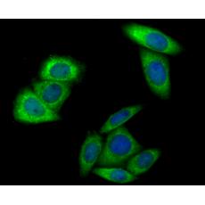
Fig1: ICC staining HKDC1 in LOVO cells (green). The nuclear counter stain is DAPI (blue). Cells were fixed in paraformaldehyde, permeabilised with 0.25% Triton X100/PBS.

Fig2: ICC staining HKDC1 in MCF-7 cells (green). The nuclear counter stain is DAPI (blue). Cells were fixed in paraformaldehyde, permeabilised with 0.25% Triton X100/PBS.

Fig3: ICC staining HKDC1 in SiHa cells (green). The nuclear counter stain is DAPI (blue). Cells were fixed in paraformaldehyde, permeabilised with 0.25% Triton X100/PBS.

Fig4: Immunohistochemical analysis of paraffin-embedded rat kidney tissue using anti-HKDC1 antibody. Counter stained with hematoxylin.

Fig5: Immunohistochemical analysis of paraffin-embedded mouse brain tissue using anti-HKDC1 antibody. Counter stained with hematoxylin.

Fig6: Flow cytometric analysis of LOVO cells with HKDC1 antibody at 1/100 dilution (red) compared with an unlabelled control (cells without incubation with primary antibody; green). Alexa Fluor 488-conjugated goat anti-rabbit IgG was used as the secondary antibody.
特别提示:本公司的所有产品仅可用于科研实验,严禁用于临床医疗及其他非科研用途!


