Anti-Cytokeratin 8 antibody
-
概述
- 产品描述Cytokeratins comprise a diverse group of intermediate filament proteins (IFPs) that are expressed as pairs in both keratinized and non-keratinized epithelial tissue. Cytokeratins play a critical role in differentiation and tissue specialization and function to maintain the overall structural integrity of epithelial cells. They have been found to be useful markers of tissue differentiation, which is directly applicable to the characterization of malignant tumors. Cytokeratin 8 expression is seen in epithelium and epithelium-derived tumors. The Cytokeratin 8 and 18 pair are normally expressed in simple epithelia, but not in stratified epithelial cells. Research indicates that squamous cell carcinomas derived from stratified epithelia show abnormal expression of Cytokeratin 8 and 18, although it is not known whether these proteins contribute to the malignant phenotype of the cells. Expression of Cytokeratin 8 and 18 in oral squamous cell carcinomas is an independent prognostic marker that indicates a poor prognosis. Cytokeratin 8 expression correlates with malignancy in leukoplakia and carcinomas of the head and neck; it is expressed in all non-small-cell lung cancers. Cytokeratin 8 has been shown to possess extracellular epitopes on tumor cells, which may represent valuable targets for therapy.
- 产品名称Anti-Cytokeratin 8 antibody
- 分子量53 kDa
- 种属反应性Human,Mouse,Rat
- 验证应用WB,ICC,IHC-P,FC
- 抗体类型兔多抗
- 免疫原Peptide
- 偶联Non-conjugated
-
性能
- 形态Liquid
- 浓度1 mg/mL.
- 存放说明Store at +4℃ after thawing. Aliquot store at -20℃ or -80℃. Avoid repeated freeze / thaw cycles.
- 存储缓冲液1*PBS (pH7.4), 0.2% BSA, 50% Glycerol. Preservative: 0.05% Sodium Azide.
- 亚型IgG
- 纯化方式Peptide affinity purified
- 亚细胞定位Cytoplasm. Nucleus
- 其它名称CARD2 antibody
CK 8 antibody
CK-8 antibody
CK8 antibody
CYK8 antibody
CYKER antibody
Cytokeratin endo A antibody
Cytokeratin-8 antibody
DreK8 antibody
EndoA antibody
K2C8 antibody
K2C8_HUMAN antibody
K8 antibody
Keratin 8 antibody
Keratin type II cytoskeletal 8 antibody
Keratin, type II cytoskeletal 8 antibody
Keratin-8 antibody
KO antibody
Krt 2.8 antibody
KRT8 antibody
MGC118110 antibody
MGC174782 antibody
MGC53564 antibody
MGC85764 antibody
sb:cb186 antibody
Type-II keratin Kb8 antibody
more
-
应用
WB: 1:500-1:1,000
ICC: 1:50-1:200
IHC-P: 1:50-1:200
FC: 1:50-1:100
-
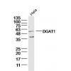
Fig1: Western blot analysis of Cytokeratin 8 on different lysates using anti-Cytokeratin 8 antibody at 1/500 dilution.
Positive control:
Lane 1: Human small intestine
Lane 2: A431
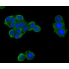
Fig2: ICC staining Cytokeratin 8 in A549 cells (green). The nuclear counter stain is DAPI (blue). Cells were fixed in paraformaldehyde, permeabilised with 0.25% Triton X100/PBS.
Fig3: ICC staining Cytokeratin 8 in LOVO cells (green). The nuclear counter stain is DAPI (blue). Cells were fixed in paraformaldehyde, permeabilised with 0.25% Triton X100/PBS.
Fig4: ICC staining Cytokeratin 8 in MCF-7 cells (green). The nuclear counter stain is DAPI (blue). Cells were fixed in paraformaldehyde, permeabilised with 0.25% Triton X100/PBS.
Fig5: Immunohistochemical analysis of paraffin-embedded rat epididymis tissue using anti-Cytokeratin 8 antibody. Counter stained with hematoxylin.
Fig6: Immunohistochemical analysis of paraffin-embedded human lung cancer tissue using anti-Cytokeratin 8 antibody. Counter stained with hematoxylin.
Fig7: Immunohistochemical analysis of paraffin-embedded human prostate tissue using anti-Cytokeratin 8 antibody. Counter stained with hematoxylin.
Fig8: Immunohistochemical analysis of paraffin-embedded mouse liver tissue using anti-Cytokeratin 8 antibody. Counter stained with hematoxylin.
Fig9: Flow cytometric analysis of MCF-7 cells with Cytokeratin 8 antibody at 1/100 dilution (red) compared with an unlabelled control (cells without incubation with primary antibody; black). Alexa Fluor 488-conjugated goat anti-rabbit IgG was used as the
特别提示:本公司的所有产品仅可用于科研实验,严禁用于临床医疗及其他非科研用途!