Anti-EPOR antibody
-
概述
- 产品描述This gene encodes the erythropoietin receptor which is a member of the cytokine receptor family. Upon erythropoietin binding, this receptor activates Jak2 tyrosine kinase which activates different intracellular pathways including: Ras/MAP kinase, phosphatidylinositol 3-kinase and STAT transcription factors. The stimulated erythropoietin receptor appears to have a role in erythroid cell survival. Defects in the erythropoietin receptor may produce erythroleukemia and familial erythrocytosis. Dysregulation of this gene may affect the growth of certain tumors. Alternate splicing results in multiple transcript variants. Receptor for erythropoietin. Mediates erythropoietin-induced erythroblast proliferation and differentiation. Upon EPO stimulation, EPOR dimerizes triggering the JAK2/STAT5 signaling cascade. In some cell types, can also activate STAT1 and STAT3. May also activate the LYN tyrosine kinase. Isoform EPOR-T acts as a dominant-negative receptor of EPOR-mediated signaling.
- 产品名称Anti-EPOR antibody
- 分子量56 KDa
- 种属反应性Human,Mouse,Rat,Dog,Cow,Horse
- 验证应用WB,IHC-P,FC
- 抗体类型兔多抗
- 免疫原KLH conjugated synthetic peptide derived from human EPOR 301-450/508
- 偶联Non-conjugated
-
性能
- 形态Liquid
- 浓度1 mg/mL.
- 存放说明Store at -20℃ for one year. Avoid repeated freeze/thaw cycles. The lyophilized antibody is stable at room temperature for at least one month and for greater than a year when kept at -20℃. When reconstituted in sterile pH 7.4 0.01M PBS or diluent of antibody the antibody is stable for at least two weeks at 2-4℃.
- 存储缓冲液0.01M TBS(pH7.4) with 1% BSA, 0.03% Proclin300 and 50% Glycerol.
- 亚型IgG
- 纯化方式affinity purified by Protein A
- 亚细胞定位Cell membrane; Single-pass type I membrane protein. Isoform EPOR-S: Secreted. Note=Secreted and located to the cell surface.
- 其它名称erythropoietin receptor
EPO R
EPO Receptor
Erythropoietin receptor precursor
EPOR_HUMAN
MGC138358.
-
应用
WB:1:500-2000
IHC-P:1:400-800
FC:1µg/Test
-
Fig1: Blank control: Molt-4 Cells(blue).
Primary Antibody: Rabbit Anti-EPOR/FITC Conjugated antibody Dilution: 1μg in 100 μL 1X PBS containing 0.5% BSA;
Isotype Control Antibody: Rabbit IgG/AF488 orange) ,used under the same conditions.
Protocol
The cells were fixed with 2% paraformaldehyde (10 min) . The cells were washed twice with 1 X PBS. The cells were incubated in 1 X PBS containing 0.5% BSA + 1 0% goat serum (15 min) to block non-specific protein-protein interactions followed by the incubated with antibody (ER1908-28/AF488, 1μg /1x10^6 cells) for 30 min on ice. Acquisition of 20,000 events was performed.
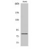
Fig2: Sample:
K562(Human) Cell Lysate at 30 ug
Primary: Anti-EPORat 1/300 dilution
Secondary: IRDye800CW Goat Anti-Rabbit IgG at 1/20000 dilution
Predicted band size: 56 kD
Observed band size: 30/52 kD
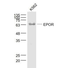
Fig3: Sample:
K562(Human) Cell Lysate at 30 ug
Primary: Anti- EPORat 1/1000 dilution
Secondary: IRDye800CW Goat Anti-Rabbit IgG at 1/20000 dilution
Predicted band size: 56 kD
Observed band size: 64 kD
特别提示:本公司的所有产品仅可用于科研实验,严禁用于临床医疗及其他非科研用途!


