Anti-LSD1 antibody
-
概述
- 产品描述Histone methylation regulates chromatin structure and transcription and maintains an epigenetic state of the cell. Histone methylation is dynamically regulated by histone methylases and demethylases. Lysine-specific histone demethylase 1 (LSD1), also designated BHC110, is a flavin-dependent amine oxidase which catalyzes the removal of one or two methyl groups from the methyl-lysine-4 side chain of Histone H3. The LSD1 protein contains a SWIRM domain, a FAD-binding motif and an amine oxidase domain. Association with CoREST, a SANT domain-containing corepressor, positively regulates LSD1. CoREST mediates the demethylation ability of LSD1 and protects it from proteasomal degradation in vivo. PHF21A (also designated BCH80), a PHD domain-containing protein, inhibits activity of LSD1/CoREST mediated de-methylation. The LSD1 protein also co-localizes with the androgen receptor in human prostate tumor cells and in unaffected prostate cells, stimulating androgen-receptor-dependent transcription.
- 产品名称Anti-LSD1 antibody
- 分子量110 kDa
- 种属反应性Human,Mouse,Rat
- 验证应用WB,ICC,IHC-P
- 抗体类型兔多抗
- 免疫原Peptide
- 偶联Non-conjugated
-
性能
- 形态Liquid
- 浓度1 mg/mL.
- 存放说明Store at +4℃ after thawing. Aliquot store at -20℃ or -80℃. Avoid repeated freeze / thaw cycles.
- 存储缓冲液1*PBS (pH7.4), 0.2% BSA, 50% Glycerol. Preservative: 0.05% Sodium Azide.
- 亚型IgG
- 纯化方式Peptide affinity purified
- 亚细胞定位Nucleus.
- 其它名称Amine oxidase (flavin containing) domain 2 antibody
Amine oxidase, flavin containing, 2 antibody
AOF2 antibody
BHC110 antibody
BRAF35 HDAC complex protein BHC110 antibody
BRAF35-HDAC complex protein BHC110 antibody
BRAF35/HDAC complex, 110-kD subunit antibody
CPRF antibody
EC1 antibody
FAD binding protein BRAF35 HDAC complex, 110 kDa subunit antibody
Flavin-containing amine oxidase domain-containing protein 2 antibody
KDM 1 antibody
KDM1 antibody
Kdm1a antibody
KDM1A_HUMAN antibody
KIAA0601 antibody
LSD 1 antibody
LSD1 antibody
Lysine (K) specific demethylase 1 antibody
Lysine (K) specific demethylase 1A antibody
Lysine demethylase 1A antibody
Lysine specific histone demethylase 1 antibody
Lysine specific histone demethylase 1A antibody
Lysine-specific demethylase 1 antibody
Lysine-specific demethylase 1A antibody
Lysine-specific histone demethylase 1A antibody
more
-
应用
WB: 1:500-1,000
ICC: 1:500-1:2,000
IHC-P: 1:50-1:200
FC: 1:50-1:100
-
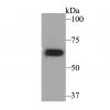
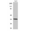
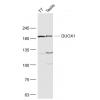
Fig1: Western blot analysis of LSD1 on PC-12 and SiHa cell lysates using anti-LSD1 antibody at 1/1,000 dilution.
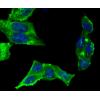
Fig2: ICC staining LSD1 in A431 cells (green). The nuclear counter stain is DAPI (blue). Cells were fixed in paraformaldehyde, permeabilised with 0.25% Triton X100/PBS.
Fig3: ICC staining LSD1 in HepG2 cells (green). The nuclear counter stain is DAPI (blue). Cells were fixed in paraformaldehyde, permeabilised with 0.25% Triton X100/PBS.
Fig4: ICC staining LSD1 in SH-SY-5Y cells (green). The nuclear counter stain is DAPI (blue). Cells were fixed in paraformaldehyde, permeabilised with 0.25% Triton X100/PBS.
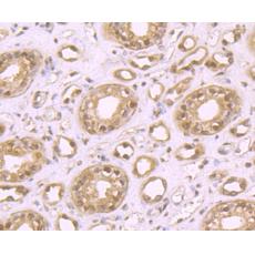
Fig5: Immunohistochemical analysis of paraffin-embedded rat kidney tissue using anti-LSD1 antibody. Counter stained with hematoxylin.
Fig6: Immunohistochemical analysis of paraffin-embedded human colon cancer tissue using anti-LSD1 antibody. Counter stained with hematoxylin.
Fig7: Immunohistochemical analysis of paraffin-embedded human spleen tissue using anti-LSD1 antibody. Counter stained with hematoxylin.
Fig8: Immunohistochemical analysis of paraffin-embedded human kidney tissue using anti-LSD1 antibody. Counter stained with hematoxylin.
Fig9: Immunohistochemical analysis of paraffin-embedded mouse cerebellum tissue using anti-LSD1 antibody. Counter stained with hematoxylin.
Fig10: Flow cytometric analysis of K562 cells with LSD1 antibody at 1/100 dilution (red) compared with an unlabelled control (cells without incubation with primary antibody; black).
特别提示:本公司的所有产品仅可用于科研实验,严禁用于临床医疗及其他非科研用途!