Anti-CD4 antibody
-
概述
- 产品描述The T cell receptor (TCR) is a heterodimer composed of either α and β or γ and δ chains. CD3 chains and the CD4 or CD8 co-receptors are also required for efficient signal transduction through the TCR. The TCR is expressed on T helper and T cytotoxic cells that can be distinguished by their expression of CD4 and CD8; T helper cells express CD4 proteins and T cytotoxic cells display CD8. CD4 is also expressed on cortical cells, mature medullary thymocytes, microglial cells and dendritic cells. CD4 (also designated T4 and Leu 3), is a membrane glycoprotein that contains four extracellular immunoglobin-like domains. The TCR in association with CD4 can bind class II MHC molecules presented by the antigen-presenting cells. The CD4 protein functions by increasing the avidity of the interaction between the TCR and an antigen-class II MHC complex. An additional role of CD4 is to function as a receptor for HIV.
- 产品名称Anti-CD4 antibody
- 分子量51 kDa
- 种属反应性Human, Mouse
- 验证应用WB,ICC,IHC-P,FC
- 抗体类型兔多抗
- 免疫原Synthetic peptide within human CD4 aa 50-100.
- 偶联Non-conjugated
-
性能
- 形态Liquid
- 浓度1 mg/mL.
- 存放说明Store at +4℃ after thawing. Aliquot store at -20℃ or -80℃. Avoid repeated freeze / thaw cycles.
- 存储缓冲液1*PBS (pH7.4), 0.2% BSA, 50% Glycerol. Preservative: 0.05% Sodium Azide.
- 亚型IgG
- 纯化方式Peptide affinity purified
- 亚细胞定位Cell membrane.
- 其它名称CD 4 antibody
CD4 (L3T4) antibody
CD4 antibody
CD4 antigen (p55) antibody
CD4 antigen antibody
CD4 molecule antibody
CD4 receptor antibody
CD4+ Lymphocyte deficiency, included antibody
CD4_HUMAN antibody
CD4mut antibody
L3T4 antibody
Leu3 antibody
Ly-4 antibody
Lymphocyte antigen CD4 antibody
MGC165891 antibody
OTTHUMP00000238897 antibody
p55 antibody
T cell antigen T4 antibody
T cell antigen T4/LEU3 antibody
T cell differentiation antigen L3T4 antibody
T cell OKT4 deficiency, included antibody
T cell surface antigen T4/Leu 3 antibody
T cell surface antigen T4/Leu3 antibody
T cell surface glycoprotein CD4 antibody
T-cell surface antigen T4/Leu-3 antibody
T-cell surface glycoprotein CD4 antibody
W3/25 antibody
W3/25 antigen antibody
more
-
应用
WB: 1:500
ICC: 1:50-1:200
IHC-P: 1:50-1:200
FC: 1:50-1:100
-
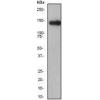
Fig1: Western blot analysis of CD4 on different tissue lysates using anti-CD4 antibody at 1/500 dilution.
Positive control:
Lane 1: Mouse spleen
Lane 2: Mouse thymus
Fig2: ICC staining CD4 in Hela cells (green). The nuclear counter stain is DAPI (blue). Cells were fixed in paraformaldehyde, permeabilised with 0.25% Triton X100/PBS.
Fig3: ICC staining CD4 in HepG2 cells (green). The nuclear counter stain is DAPI (blue). Cells were fixed in paraformaldehyde, permeabilised with 0.25% Triton X100/PBS.
Fig4: ICC staining CD4 in SH-SY5Y cells (green). The nuclear counter stain is DAPI (blue). Cells were fixed in paraformaldehyde, permeabilised with 0.25% Triton X100/PBS.
Fig5: Immunohistochemical analysis of paraffin-embedded human tonsil tissue using anti-CD4 antibody. Counter stained with hematoxylin.
Fig6: Immunohistochemical analysis of paraffin-embedded human spleen tissue using anti-CD4 antibody. Counter stained with hematoxylin.
Fig7: Immunohistochemical analysis of paraffin-embedded mouse colon tissue using anti-CD4 antibody. Counter stained with hematoxylin.
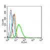
Fig8: Flow cytometric analysis of Jurkat cells with CD4 antibody at 1/100 dilution (red) compared with an unlabelled control (cells without incubation with primary antibody; black). Alexa Fluor 488-conjugated Goat anti rabbit IgG was used as the secondary
特别提示:本公司的所有产品仅可用于科研实验,严禁用于临床医疗及其他非科研用途!