Anti-CD71 antibody
-
概述
- 产品描述CD71, also known as the transferrin receptor (TFR), is a type II membrane glycoprotein that exists as a disulfide-linked homodimer of two identical subunits. CD71 binds to two molecules of transferrin and a serum iron-transport protein, and directs the cellular uptake of iron via receptor-mediated endocytosis. CD71 is expressed, typically at high levels, on all proliferating cells, reticulocytes and erythroid precursors. It is not expressed on resting leukocytes, but is upregulated upon activation of lymphocytes, monocytes and macrophages. CD71 is also found on most dividing cells and on brain endothelium. A second transferrin receptor, TFR2, also mediates the uptake of transferrin-bound iron. TFR2 is a two-subunit homodimer and is highly expressed in liver as well as in hepatocytes and erythroid precursors. Mutations in the TFR2 gene result in hereditary hemochromatosis type III (HFE3), an iron overloading disorder predominant in Caucasians.
- 产品名称Anti-CD71 antibody
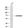
- 分子量84 kDa
- 种属反应性Human,Mouse
- 验证应用ICC,IHC-P,FC
- 抗体类型兔多抗
- 免疫原Recombinant protein
- 偶联Non-conjugated
-
性能
- 形态Liquid
- 浓度1 mg/mL.
- 存放说明Store at +4℃ after thawing. Aliquot store at -20℃ or -80℃. Avoid repeated freeze / thaw cycles.
- 存储缓冲液1*PBS (pH7.4), 0.2% BSA, 50% Glycerol. Preservative: 0.05% Sodium Azide.
- 亚型IgG
- 纯化方式Protein A purified.
- 亚细胞定位Secreted and Cell membrane. Melanosome.
- 其它名称CD 71 antibody
CD71 antibody
CD71 antigen antibody
IMD46 antibody
OTTHUMP00000208523 antibody
OTTHUMP00000208524 antibody
OTTHUMP00000208525 antibody
p90 antibody
sTfR antibody
T9 antibody
TFR 1 antibody
TfR antibody
TfR1 antibody
TFR1_HUMAN antibody
TFRC antibody
TR antibody
Transferrin receptor (p90 CD71) antibody
Transferrin receptor protein 1, serum form antibody
Trfr antibody
more
-
应用
ICC: 1:50-1:200
IHC-P: 1:50-1:200
FC: 1:50-1:100
-
Fig1: ICC staining CD71 in PC-3M cells (green). The nuclear counter stain is DAPI (blue). Cells were fixed in paraformaldehyde, permeabilised with 0.25% Triton X100/PBS.
Fig2: ICC staining CD71 in A549 cells (green). The nuclear counter stain is DAPI (blue). Cells were fixed in paraformaldehyde, permeabilised with 0.25% Triton X100/PBS.
Fig3: ICC staining CD71 in MCF-7 cells (green). The nuclear counter stain is DAPI (blue). Cells were fixed in paraformaldehyde, permeabilised with 0.25% Triton X100/PBS.
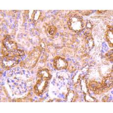
Fig4: Immunohistochemical analysis of paraffin-embedded mouse kidney tissue using anti-CD71 antibody. Counter stained with hematoxylin.
Fig5: Immunohistochemical analysis of paraffin-embedded human kidney tissue using anti-CD71 antibody. Counter stained with hematoxylin.
Fig6: Immunohistochemical analysis of paraffin-embedded human placenta tissue using anti-CD71 antibody. Counter stained with hematoxylin.
Fig7: Immunohistochemical analysis of paraffin-embedded mouse brain tissue using anti-CD71 antibody. Counter stained with hematoxylin.
Fig8: Flow cytometric analysis of HL-60 cells with CD71 antibody at 1/100 dilution (red) compared with an unlabelled control (cells without incubation with primary antibody; black). Alexa Fluor 488-conjugated Goat anti rabbit IgG was used as the secondary
特别提示:本公司的所有产品仅可用于科研实验,严禁用于临床医疗及其他非科研用途!