Anti-DKK1 antibody
-
概述
- 产品描述The Wnt genes are a group of well conserved, cysteine-rich secreted glycoproteins that are required for numerous developmental processes including embryogenesis, asymmetric cell division and central nervous system (CNS) patterning. Wnt association with the seven membrane spanning receptor frizzled activates dishevelled, which downregulates glycogen synthase kinase (GSK) through serine phosphorylation, causing the accumulation of b-catenin and subsequent regulation of developmentally significant Wnt |target genes. The Dickkopf family of secreted inhibitors of Wnt signaling ensures proper morphological development by antagonizing different stages of the Wnt cascade. Dkk-1 (Dickkopf-1) acts upstream of b-catenin and dishevelled to inhibit Wnt signaling. Dkk-1 is a 266-amino acid (human), secreted protein that contains a 31-residue N-terminal signal peptide, 2 cysteine rich domains, and a putative carboxy terminal N-glycosylation site. Human Dkk-1 transcripts are abundantly present in fetal kidney, adult placenta and adult prostate. Putative cis regulatory elements upstream of the Dkk-1 start site include p53, Sp1, MyoD, STAT, Oct-1/2, C/EBP-a, C/EBP-b, GATA-1, GATA-2 and GATA-3.
- 产品名称Anti-DKK1 antibody
- 分子量26 kDa
- 种属反应性Human,Mouse
- 验证应用WB,ICC,IHC-P,FC
- 抗体类型兔多抗
- 免疫原Peptide
- 偶联Non-conjugated
-
性能
- 形态Liquid
- 浓度1 mg/mL.
- 存放说明Store at +4℃ after thawing. Aliquot store at -20℃ or -80℃. Avoid repeated freeze / thaw cycles.
- 存储缓冲液1*PBS (pH7.4), 0.2% BSA, 40% Glycerol. Preservative: 0.05% Sodium Azide.
- 亚型IgG
- 纯化方式Peptide affinity purified
- 亚细胞定位Secreted.
- 其它名称Dickkopf 1 antibody
Dickkopf 1 homolog antibody
Dickkopf 1 like antibody
Dickkopf homolog 1 antibody
Dickkopf like protein 1 antibody
Dickkopf related protein 1 antibody
Dickkopf WNT signaling pathway inhibitor 1 antibody
Dickkopf-1 antibody
Dickkopf-related protein 1 antibody
DKK 1 antibody
Dkk-1 antibody
Dkk1 antibody
DKK1_HUMAN antibody
hDkk 1 antibody
hDkk-1 antibody
SK antibody
more
-
应用
WB: 1:500
ICC: 1:50-1:200
IHC-P: 1:50-1:200
FC: 1:50-1:100
-
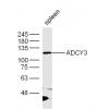
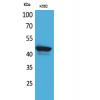
Fig1: Western blot analysis of DKK1 on different tissue lysates using anti-DKK1 antibody at 1/500 dilution.
Positive control:
Lane 1: Mouse brain
Lane 2: Human fetal brain
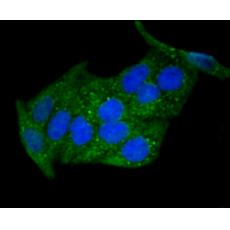
Fig2: ICC staining DKK1 in Hela cells (green). The nuclear counter stain is DAPI (blue). Cells were fixed in paraformaldehyde, permeabilised with 0.25% Triton X100/PBS.
Fig3: ICC staining DKK1 in NCCIT cells (green). The nuclear counter stain is DAPI (blue). Cells were fixed in paraformaldehyde, permeabilised with 0.25% Triton X100/PBS.
Fig4: Immunohistochemical analysis of paraffin-embedded human placenta tissue using anti-DKK1 antibody. Counter stained with hematoxylin.
Fig5: Immunohistochemical analysis of paraffin-embedded mouse placenta tissue using anti-DKK1 antibody. Counter stained with hematoxylin.
特别提示:本公司的所有产品仅可用于科研实验,严禁用于临床医疗及其他非科研用途!